Overview
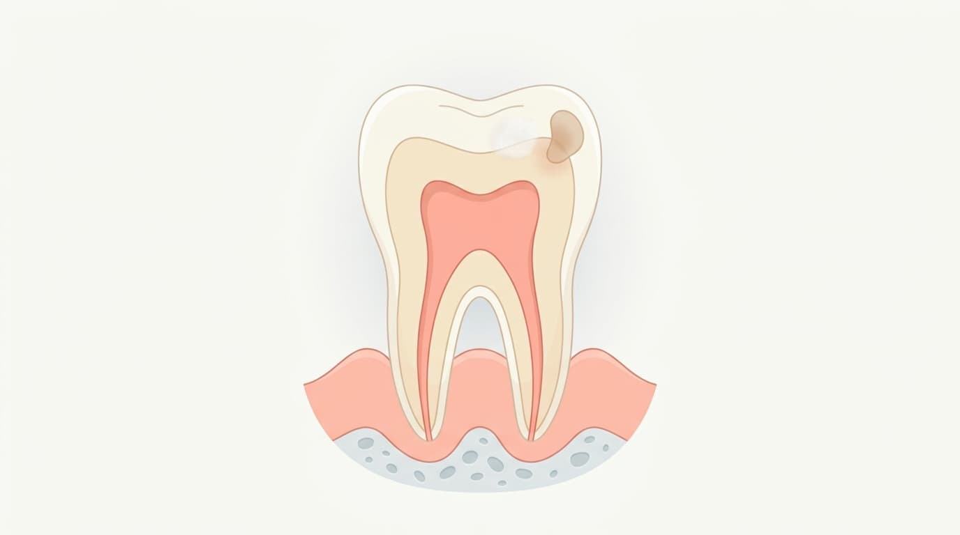
Dental caries (tooth decay) is a disease in which acids produced when bacteria in the dental plaque on the tooth surface break down sugars from food dissolve the minerals on the outer tooth surface, gradually destroying tooth structure. The lesion may progress or arrest depending on the balance between four factors (bacteria, fermentable carbohydrates, the tooth, and time) and protective factors such as the buffering action of saliva and fluoride. When the acidity of the plaque drops below pH 5.5, the minerals that make up enamel begin to dissolve, and if this process continues it leads to a visible hole (cavity).
Causes
The most common cause is the process by which bacteria such as Streptococcus mutans in dental plaque break down sugar (sucrose) to produce acid. Eating sweets frequently or brushing insufficiently lengthens the time acid is produced, making the lesion more likely to progress.
One important cause is hyposalivation (dry mouth). When saliva flow drops sharply due to medications such as antidepressants or diuretics, radiation therapy, or Sjögren's syndrome, saliva's self-cleansing and buffering actions are lost, and decay can spread explosively across multiple teeth in a short period.
Symptoms
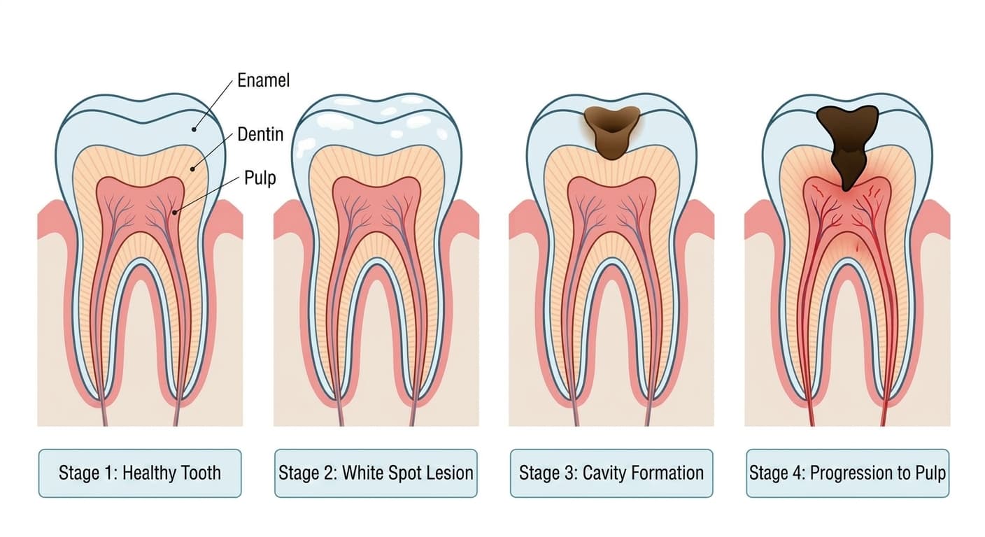
In the early stage, the tooth surface often just shows a hazy white spot (white spot lesion) with no pain. As it progresses, the spot turns brown or black, an actual hole forms on the surface, and sensitivity to cold water or sweet food may appear.
That said, even a brown or black lesion may be an arrested, inactive lesion if it is hard, smooth, and glossy, so factors besides color need to be considered together.
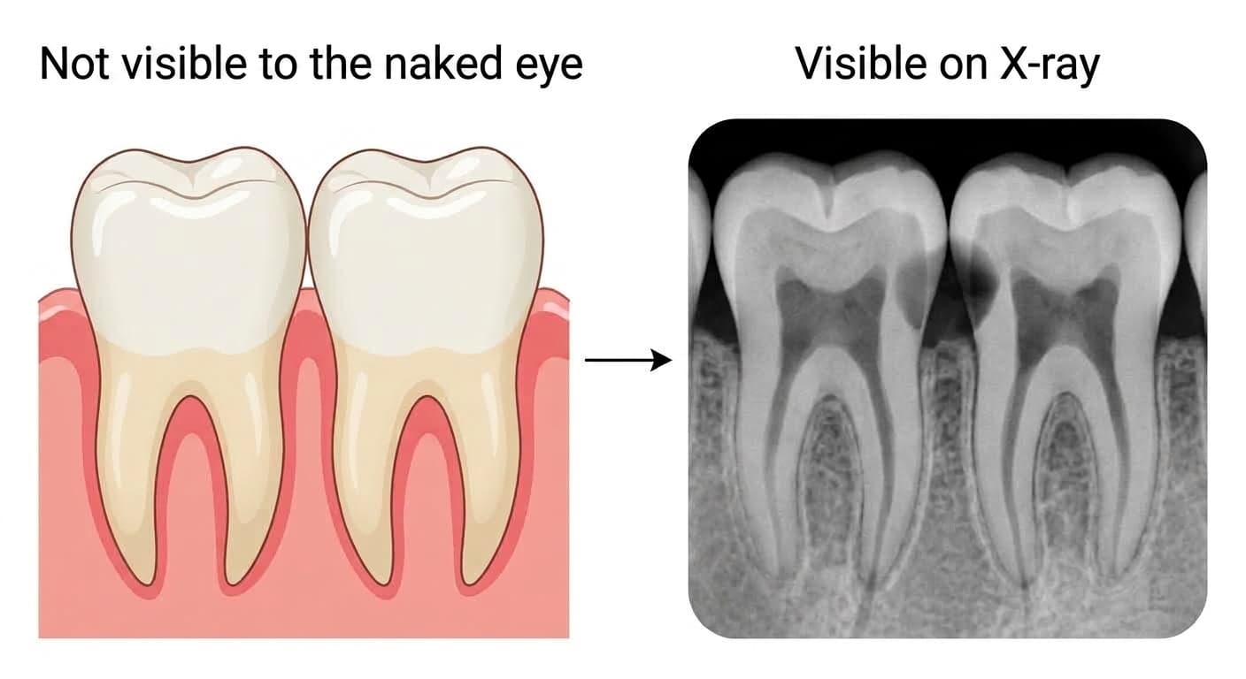
There are exceptions to this as well. Early decay that forms between adjacent teeth (the proximal surface) is not visible to the naked eye and is difficult to confirm even by scraping it with an instrument, so it commonly progresses quite far with no symptoms at all. The absence of symptoms does not mean the absence of decay.
Diagnosis
Diagnosis starts with asking about the symptoms and examining the tooth surface by eye and with an instrument, combined with radiographic examination to find lesions that are not visible. X-rays are especially useful because proximal (between-teeth) decay and new decay beneath an existing filling are difficult to confirm by sight or with an instrument alone. These images allow the direction and extent of the lesion to be assessed, so people prone to decay have them taken every 3 to 6 months as a reference for deciding whether treatment is needed. However, pressing too hard on a weakened surface with an instrument can actually break it, so diagnosis is not based on a single test but on a combination of findings.
Treatment & Where to Go
At the white spot stage, before a cavity has formed, applying remineralizing agents such as fluoride without any removal of tooth structure, along with managing brushing and diet and monitoring the lesion, may be enough to arrest or reverse it. Once a cavity has formed and needs to be filled, the standard approach is a minimally invasive one: selectively removing only the bacteria-infected dentin, preserving as much healthy tooth structure as possible, and then filling with resin or a similar material. Decay that is deep enough to be close to the pulp (nerve) requires an additional pulp-protecting procedure. If bacteria spread to the pulp and cause irreversible inflammation, root canal treatment is needed, and if destruction is too extensive for the tooth to be saved, extraction may even be considered. Care is provided by a dentist who treats cavities and performs root canal treatment.
Outlook & Precautions
If left untreated, bacteria and their toxins can travel through tiny channels in the dentin (dentinal tubules) to the pulp (nerve) and cause inflammation, and if it progresses further, the nerve can die (pulp necrosis) or inflammation can spread around the tooth root. After treatment, regular checkups and X-rays every 3 to 6 months, and fluoride applications every 3 months for those at high risk of decay, help prevent recurrence. Sensitivity to cold water or sweets usually subsides once the cause is removed, but a throbbing pain that appears on its own without any stimulus may be a sign that the nerve has been affected, and prompt care should be sought.