
Overview
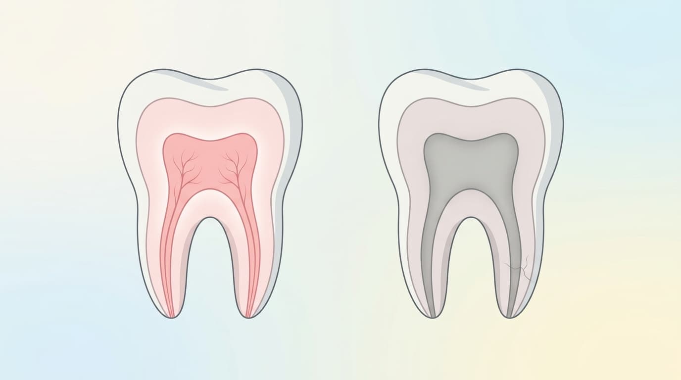
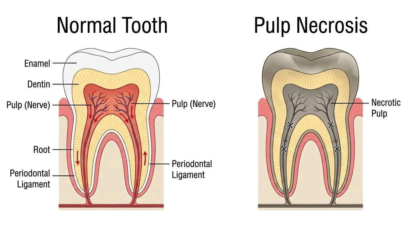
Pulp necrosis refers to a condition in which blood circulation to the pulp (the nerve and blood vessel tissue inside the tooth, commonly called the "tooth nerve") stops completely, causing the entire tissue to die. When the supply of oxygen and nutrients to the pulp is cut off by bacterial infection, trauma, deep dental caries, or other causes, the tissue does not recover and dies. Once the pulp has died, it no longer responds to cold, heat, or electrical stimulation, which is why pain often disappears rather than continuing. However, the absence of pain does not mean the tooth has healed; it can be a sign that bacteria are continuing to grow inside the dead tissue.
Causes
The most common cause is deep dental caries (tooth decay) that progresses down to the nerve and infects the pulp. A typical pathway is neglected irreversible pulpitis (nerve inflammation that does not resolve even after the irritant is removed) progressing over time into pulp necrosis. Trauma such as a cracked or knocked tooth that cuts off blood flow to the pulp is also a common cause.
Less common but important causes include cracks that form in the tooth, exposure of dentinal tubules on the tooth surface that allows bacteria to penetrate, and severe periodontal disease that cuts off the blood supply from the root side of the tooth. These cases are easy to miss because the pulp can die even when there is no obvious decay or trauma visible from the outside.
Symptoms
The most distinctive symptom is, paradoxically, the absence of symptoms. Because the dead nerve can no longer transmit sensation, the tooth often does not respond to cold water or hot food, and pain that was present may pass without warning at some point. A noticeable sign can be a darkening of the tooth color, turning gray, brown, or in severe cases a dark blue-gray, as blood breakdown products from inside the tooth seep into the dentin.
However, not every case is symptom-free. If the inflammation spreads to the tissue around the root tip, chewing or tapping on the tooth can cause pain, and the gums may swell and discharge pus. Even without pain, you should not assume everything is fine. If the tooth's color has changed or you feel discomfort while chewing, you should see a dentist.
Diagnosis
Pulp necrosis is diagnosed by combining several tests. Applying a mild electrical stimulus and applying cold and hot water to the tooth are the basic ways to check whether a living nerve is present; if the pulp has died, there is no response to either test. However, because partial necrosis or testing conditions can sometimes produce a response in a dead nerve or no response in a living one, diagnosis is not based on a single test result alone.
Tapping the tooth to check for inflammation around the root, pressing on the gum with a finger to check for swelling or pus, and radiographs to examine changes in the bone around the root tip are used together. If these tests are still not conclusive, the dentist may lightly cut into the tooth without anesthesia to check whether any sensation remains, and in complex cases a three-dimensional CT scan is used to precisely examine the extent of the lesion.
Treatment & Where to Go
The standard treatment for pulp necrosis is root canal treatment (nerve treatment). This involves removing the dead pulp tissue and the bacteria within it, cleaning and disinfecting the inside of the root canal, then filling and sealing it, performed in the conservative dentistry (endodontics) department. If an acute abscess has formed at the root tip and the gum is swollen and severely painful, an incision may first be made to drain the pus before root canal treatment proceeds, or acute drainage may instead be performed by cutting into the tooth and draining through the root canal.
If non-surgical root canal treatment fails to heal the lesion, or if the shape of the root canal makes a non-surgical approach difficult, surgical root canal treatment may be considered. This involves directly opening the area around the root tip to remove the infected tissue and a portion of the root end, then filling it from the reverse direction; success rates have improved in recent years with the use of surgical microscopes and biocompatible materials. At every stage, care from a dentist who performs root canal treatment is needed.
Outlook & Precautions
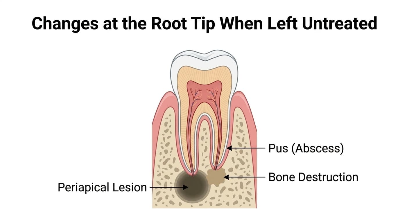
If left untreated, bacteria and toxins inside the dead pulp can spread beyond the root tip, progressing into conditions such as apical periodontitis, acute or chronic periapical (alveolar) abscess, granuloma, or a periapical cyst. Even without symptoms, the bone around the root tip can be gradually destroyed while the condition is left untreated.
You should see a dentist right away in the following situations: if the gum suddenly swells or becomes inflamed, if swelling accompanied by heat spreads to the face or neck, or if there is unbearable pain while chewing. After root canal treatment, follow-up monitoring is needed to regularly check whether pain on tapping has decreased and whether the lesion at the root tip is shrinking on radiographs.