

Question
There are cases where a patient notices a dark, empty-looking area at the root of a tooth on an X-ray taken to check progress after finishing root canal treatment, and worries about it. This addresses the concern about whether it is possible to tell from the image alone whether the treatment went wrong and whether the tooth needs to be retreated.
Short Answer
A single X-ray image showing a gap is not enough on its own to determine whether retreatment is needed. First, it must be determined whether the gap is a void inside the filling material or a radiolucency remaining around the root apex, and in both cases it is necessary to check whether there are symptoms such as pain or swelling, and whether the area narrows or grows larger over time. An area that looked fine right after treatment can temporarily remain visible during the healing process, and conversely, an area can be healing well without symptoms even though it does not completely disappear on the image. So the key to judgment is following how the area changes over time rather than relying on a single image.
Why Is This the Case?
When a Gap Remains Inside the Filling Material
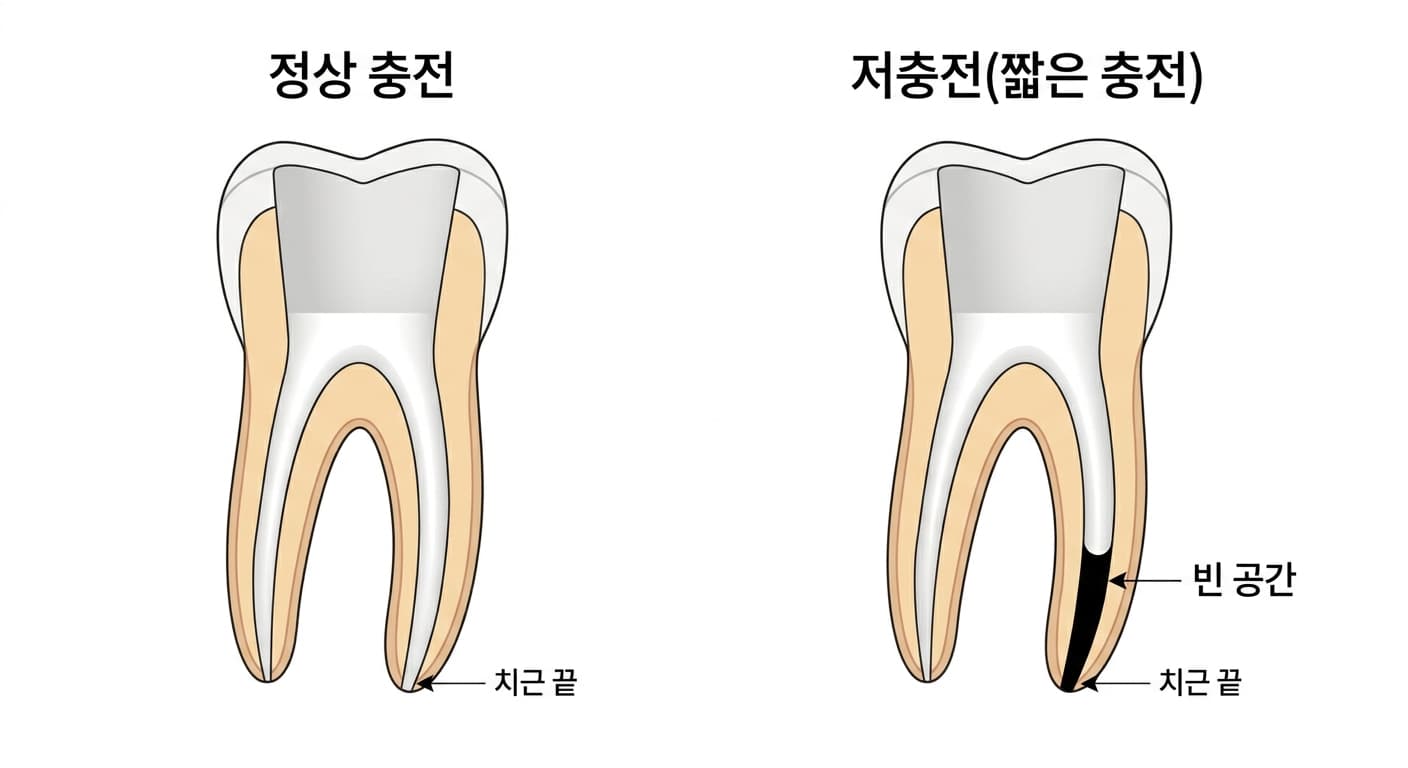
The final step of root canal treatment is root canal filling (obturation), which completely fills the space inside the root, from which bacteria have been removed, with filling material. The purpose of this filling is not simply to fill a hole; it is to block, from the apical, lateral, and coronal directions alike, the pathway through which bacteria and tissue fluid could move between the oral cavity or periapical tissue and the canal. Sealing off every possible leak like this is called sealing, and one study in the United States (the Washington study) reported that 58.66% of root canal treatment failures stemmed from cases where this seal was inadequate.
If the filling material stops short of reaching the narrowing point at the root apex, tissue fluid can pool in the empty space below it, creating a place for bacteria to grow again. However, if the end of the filling material is within 0mm to 2mm of the root apex, it is considered an adequate fill by American Association of Endodontists standards. In other words, the fact that the filling on an X-ray does not reach exactly to the root apex does not in itself mean the treatment failed. In fact, cases have been reported of teeth with a short root canal filling remaining free of notable symptoms even in images taken five years later. Conversely, if a short filling is accompanied by pain, swelling, or a periapical lesion that newly appears or grows larger, this is taken as a sign that the seal was in fact inadequate.
A Dark Area Around the Root Apex May Be a Sign of Healing
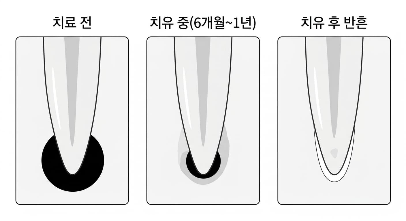
If bacteria inside a tooth spread to the tissue around the root apex before root canal treatment, inflammation called apical periodontitis develops, and the bone in that area is resorbed, appearing dark on an X-ray. Once root canal treatment removes the source of infection, this tissue does not recover immediately; it is gradually filled in with new bone, and complete healing takes 6 months to 1 year. If a new X-ray is taken during this period, the dark area can be seen gradually growing lighter, and the bone pattern (trabeculae) along with the thin white line surrounding the root (the lamina dura) becomes distinct again.
However, if a lesion was large enough to have perforated through both the buccal and lingual (cheek-side and tongue-side) bone, the area can finish healing filled with fibrous connective tissue instead of new bone. In this case, even though the tissue has already stabilized, a radiolucency continues to appear on the X-ray, looking like a scar. In fact, cases have been reported where, even five years after treating a large apical lesion, the radiolucency at the apex had not completely disappeared, yet there were no symptoms or other signs, and the border around the root was distinct. Because such a scar can look similar in appearance to a case where infection remains in the canal and is progressing toward failure, care is needed not to distinguish it by the image alone and undergo unnecessary retreatment.
What Determines Whether Retreatment Is Needed
For this reason, when determining whether treatment has failed, several factors are considered together rather than a single X-ray image. The decision takes into account the treatment history and changes in symptoms, clinical findings such as percussion testing (lightly tapping the tooth to check the response), changes in imaging over time such as whether a lesion has grown larger or smaller compared to previous images, and even the condition of the restoration or the gums. Even after all this, the result does not necessarily lead to retreatment. If there are no symptoms and the condition remains stable, the case may simply be monitored over a period of time; if a gap inside the filling material is confirmed to be the actual cause, non-surgical retreatment, which removes the existing filling material and refills the canal, may be considered; and if access inside the canal is difficult or an actual lesion remains that is not a scar, this can lead to surgical treatment.
So if you notice a gap on an X-ray, rather than drawing your own conclusion from that single image, it leads to a more accurate judgment to check with your dentist whether you currently have symptoms and, if a previous image exists, how the area has changed compared to that image.