

Question
A question from someone who, after injuring a tooth and having an X-ray taken at the dentist's office, wants to know whether a single image can definitively show if the root is fractured. They're also curious what symptoms would reveal a root fracture.
Short Answer
A single image is often not enough to confirm it. If the root is fractured at an oblique angle, the fracture line can overlap with other structures and stay hidden depending on the shooting angle, so several images taken from different angles are compared together. A fractured root also doesn't always make the tooth mobile, and the treatment approach depends on where the fracture is and how much mobility is present.
Why Is That?
Why the Fracture Line Appears or Disappears Depending on the Angle
A root fracture can run nearly horizontal, but it can also split at an oblique angle. An oblique fracture may not be visible on a single radiograph because of differences in the shooting angle. For this reason, diagnosis is made by taking 2-3 images at different vertical angles, such as 45°, 90°, and 110°, and comparing them.
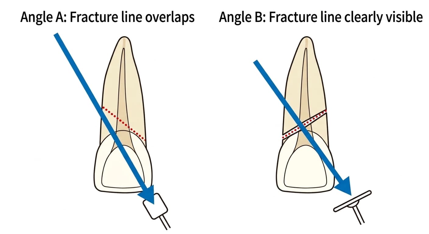
At one angle, the fracture line may overlap with tooth structures and be hard to see, while at another angle it shows up as a clear gap. If the diagnosis is still unclear even after taking images from multiple angles, a CBCT scan, which can show three-dimensional cross-sections, may be added. Even a horizontal root fracture that's hard to confirm on a plain intraoral radiograph can be seen more clearly on three-dimensional CBCT images.
What Mobility Can Tell You
Along with the images, the dentist checks the degree of mobility by gently moving the tooth with a finger or an instrument. This mobility is graded: Grade 1 if it's barely perceptible in the lip-tongue direction, Grade 2 if it moves up to about 1mm in multiple directions, and Grade 3 if it moves more than that or the tooth can be depressed or rotated.
A root fracture doesn't always produce this kind of mobility.
Why Treatment Depends on the Fracture's Location
If the fracture is in the apical third, near the root tip, and there's no mobility, it's often just monitored without any specific treatment. Conversely, if the fracture line is closer to the crown and the coronal fragment is mobile, the tooth is repositioned and then splinted together with the adjacent teeth.
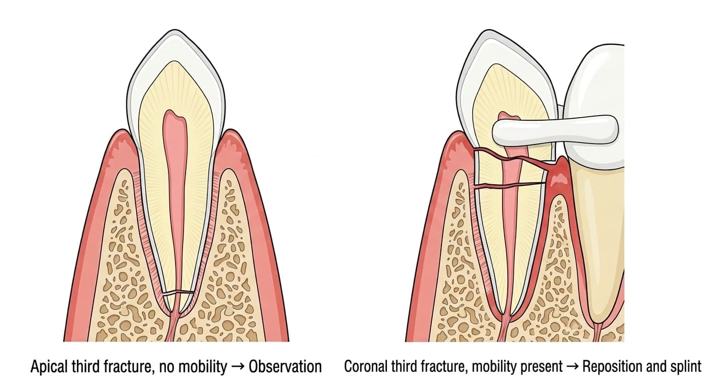
There's more than one way the fracture surfaces can heal. In some cases, calcified tissue, connective tissue, or a combination of bone and connective tissue fills in between the fracture surfaces and joins them. However, if only connective tissue forms without full union, the pulp (nerve) in the coronal fragment alone may become necrotic. In that case, rather than treating the whole tooth, only the coronal fragment's canal is filled with calcium hydroxide or MTA (a dental mineral-based filling material), root canal treatment is carried out there, and the apical fragment is left untouched.
So checking whether a fracture is present on the images is just as important as assessing its exact location and the degree of mobility together, and both are essential in determining the direction of treatment.