
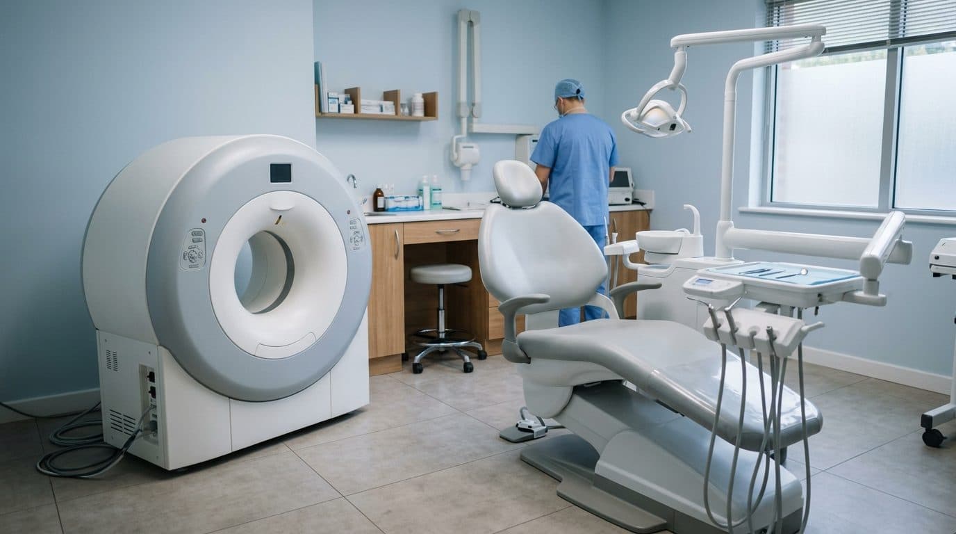
An imaging device that lets dentists examine gum inflammation, the jaw joint, and tissue around nerves without any radiation exposure has received its first formal approval in the United States.
According to dental trade outlet Orthodontic Products, the MAGNETOM Free.Max Dental Edition, a magnetic resonance imaging (MRI) system built jointly by Dentsply Sirona, the world's largest dental equipment company, and medical imaging company Siemens Healthineers, received clearance from the US Food and Drug Administration (FDA) in March. The outlet reported that this marks the first time an MRI system designed exclusively for dental use has received formal clearance in the United States.
Radiation-Free Imaging of the Teeth and Jaw Only
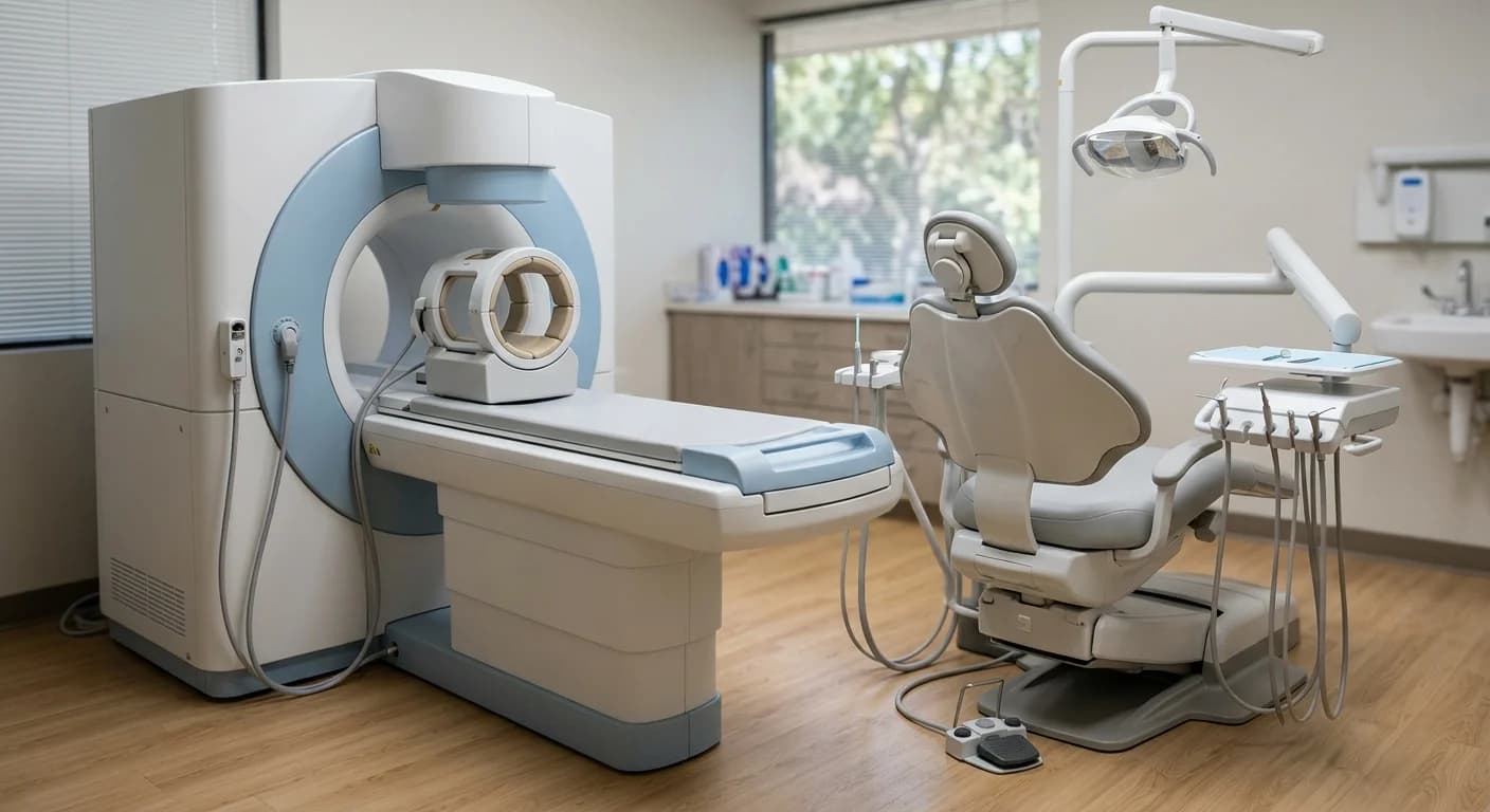
Unlike a general-purpose MRI, this system uses a dental-specific coil and a narrowed field of view, so it excludes brain tissue and scans only the teeth, jawbone, and surrounding soft tissue. The average scan takes under 20 minutes. Unlike computed tomography (CT), which uses radiation, or cone-beam CT (CBCT, the three-dimensional dental X-ray commonly used in dental offices), this system can be used repeatedly without radiation exposure concerns. That makes it especially useful for tracking a growing child's jaw joint over multiple visits or rechecking orthodontic treatment progress partway through care.
Clinical Trials in Five Areas and Diagnostic Uses
Clinical trials were conducted across five areas: orthodontics, endodontics (root canal treatment), periodontics, the temporomandibular joint, and tooth extraction. The system can distinguish between actively inflamed tissue and tissue that has already healed into scar tissue, and it can be used alongside existing tests to noninvasively assess whether a tooth's nerve (the pulp) is still alive before root canal treatment. Before extracting a wisdom tooth, it can also show in advance how close the root sits to a neighboring tooth or nerve canal. In the temporomandibular joint exams covered in What Gets Examined When Your Jaw Hurts and Won't Open Fully, the system's advantage, as the makers describe it, is that it can directly image the joint disc or inflamed areas that X-rays have long struggled to show clearly.
Yonsei University Dental Hospital's MRI and Korean Launch Plans
This is not South Korea's first encounter with this kind of imaging. Yonsei University Dental Hospital became the first dental university hospital in South Korea to bring in a dedicated 3.0T dental MRI in 2019, and has since performed more than 4,000 scans with it. It has been used to examine areas that were hard to assess with existing imaging equipment, such as disorders of the masticatory muscles, salivary glands, and tongue, and problems with the temporomandibular joint disc. The difference is that this earlier system was a large, hospital-grade general-purpose MRI repurposed for dental use, while the product that just received FDA clearance was designed from the outset as a compact system for hospitals, large dental clinics, and dental schools.
Both Dentsply Sirona and Siemens Healthineers have subsidiaries in South Korea. Dentsply Sirona Korea's website already lists its imaging equipment lineup, but plans and pricing for a South Korean launch of this dental-only MRI have not yet been disclosed. In the United States, too, pricing has not been made public, and the system is expected to roll out first to hospitals and universities, so it will likely be some time before patients can get this scan at a neighborhood dental clinic.