
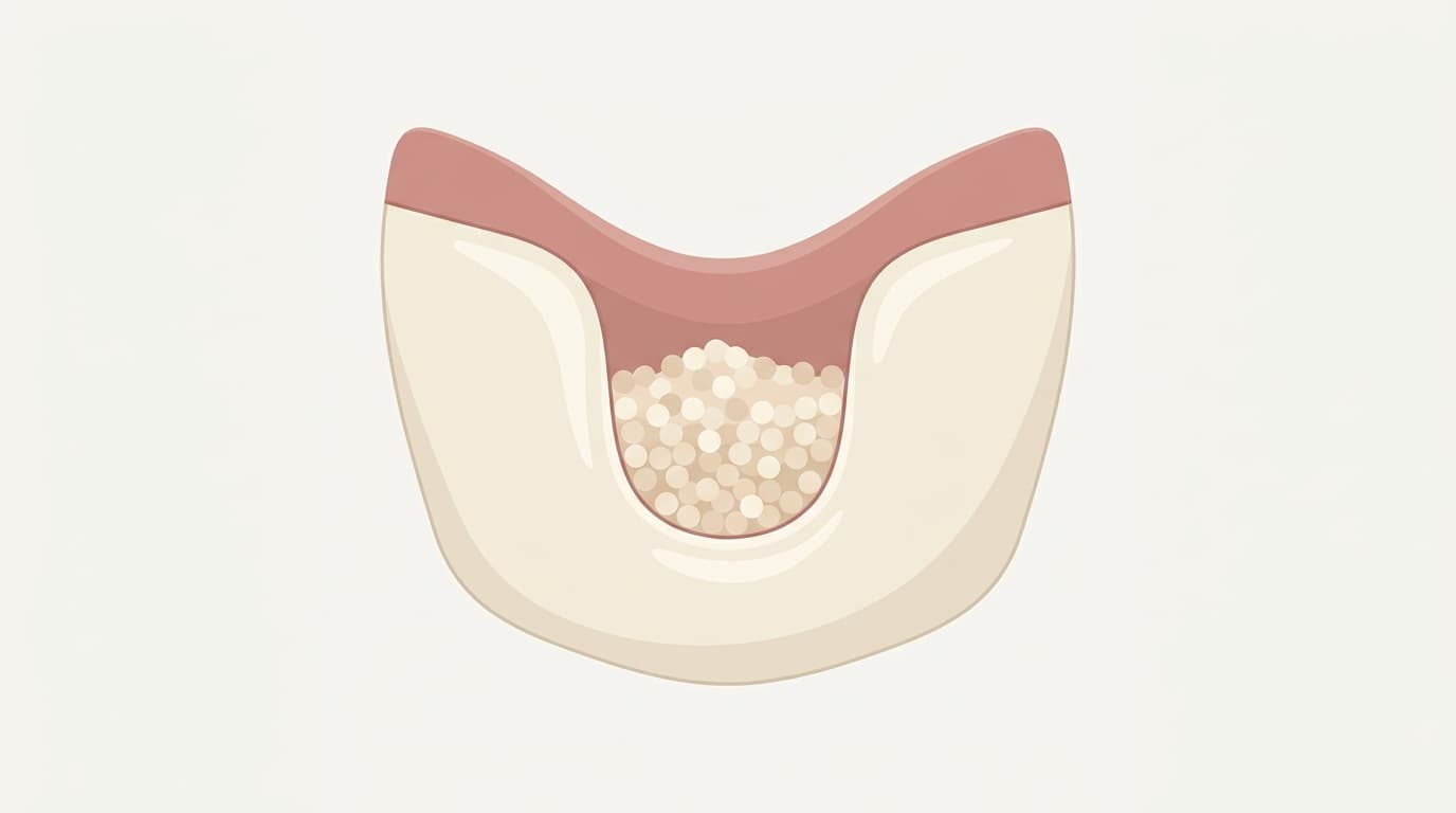
When a tooth is extracted, the alveolar bone (the jawbone that surrounds and supports the tooth) that once held it in place gradually resorbs. The buccal wall is thin and shrinks faster than other areas, and after several months in that state, the width and height available for a future implant can become insufficient. To prevent this, a treatment called alveolar ridge preservation fills the empty socket with bone graft material immediately after extraction. A recently published clinical trial directly tested whether the results are the same without the collagen barrier membrane commonly used alongside this treatment.
Why Bone Graft Is Often Covered With a Membrane
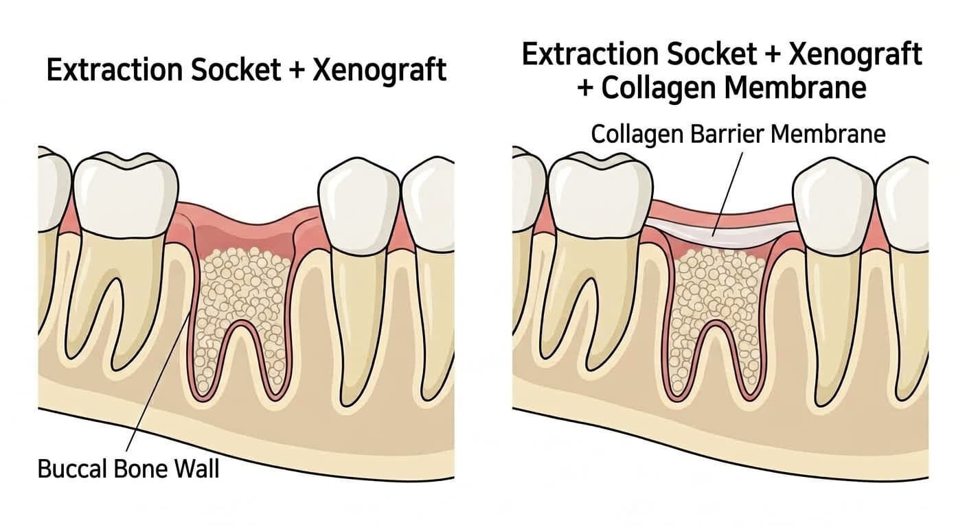
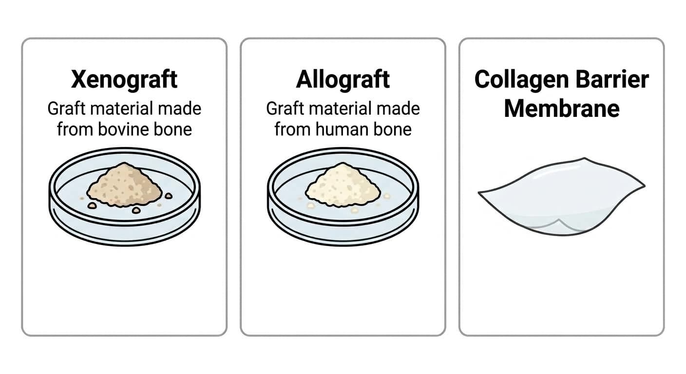
The material commonly used in alveolar ridge preservation is xenograft, made by stripping the organic components from bovine bone and leaving only the mineral. Each particle serves as a scaffold for new bone to grow on and is gradually replaced by the patient's own bone. Adding a collagen barrier membrane on top is also widely practiced. The rationale is that it stops the soft gum tissue, which grows faster than bone, from moving in and occupying the graft site first. However, adding a membrane increases material cost and procedure time, and carries its own risk of complications such as membrane exposure or infection.
After Following 20 Patients for Six Months, the Results Were the Same With or Without the Membrane
In 2026, a research team at Harvard School of Dental Medicine directly tested whether this membrane is actually necessary. They randomly divided 20 patients who had a single-rooted tooth extracted, such as a premolar, canine, or incisor, into a group that received xenograft alone and a group that received xenograft plus a collagen membrane, let the gum tissue heal on its own without forcing closure, and compared the results after six months.
| Measure (at 6 months) | Xenograft Only | Xenograft + Collagen Membrane |
|---|---|---|
| Bone width (3mm below the ridge crest) | 9.02mm | 8.41mm |
| Vertical bone height change | -0.78mm | -1.31mm |
| Keratinized gingiva width | 4.80mm | 4.80mm |
| New bone formation rate (biopsy) | 25.8% | 27.9% |
None of the four measures showed a statistically significant difference between the two groups. Given that baseline buccal bone thickness was similar in both groups, around 1mm, this suggests that at sites where some buccal bone remains, bone width is maintained at a similar level even without a membrane. The research team stated that omitting the membrane can achieve similar results while reducing material cost and procedure time. However, since this was a small trial involving only 20 patients, whether the same results hold in a larger patient population remains to be confirmed.
Other Studies Found Different Answers
This does not mean that adding material is always pointless. In a 2026 study by a research team at the University of Texas School of Dentistry that divided 120 patients into two groups, adding xenograft to human-derived allograft actually resulted in a lower rate of new bone confirmed by biopsy than using allograft alone (23-24% versus 37-41%). This echoes the Harvard finding that adding material does not always improve results, but here the difference showed up not in bone width but in the proportion of newly formed native bone.
This should not be broadly read as meaning grafting and augmentation are unnecessary altogether. The picture was different at multi-rooted molar sites with wide extraction sockets. In a 2025 study by a Brazilian research team looking at molar extraction sites, the groups that received bone graft or a titanium membrane preserved bone height better than the group left to heal spontaneously with no treatment. In the untreated group, more than two-thirds of cases saw the site subside enough to reach the maxillary sinus, requiring the use of short implants. Results from single-rooted incisors and premolars cannot simply be applied to multi-rooted molars.
A 2025 study that pooled and re-examined results from multiple clinical trials likewise found that the combination of xenograft plus membrane produced a lower rate of new bone formation than spontaneous healing with no treatment at all, whereas platelet concentrates drawn from blood produced more new bone growth than spontaneous healing. This suggests that outcomes vary widely depending on the type of bone graft material used, and that the choice of material itself may be a bigger variable in the outcome than whether or not a membrane is applied.
What this Harvard study shows is that at incisor and premolar extraction sites with some buccal bone remaining, xenograft alone can preserve bone width to a level comparable to adding a collagen membrane. Whether this finding can be extended to multi-rooted molars or sites with extensive bone damage remains a question for future research to confirm.