When the Pulp Dies in a Tooth Whose Root Has Not Finished Forming
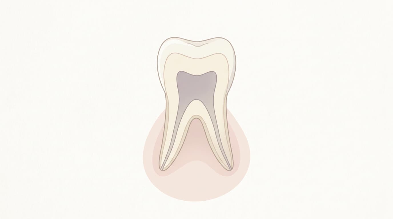
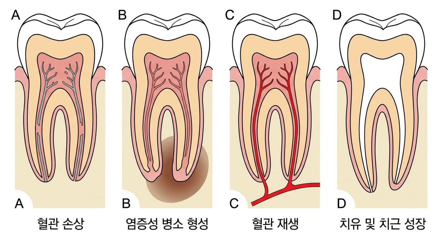
A permanent tooth that has erupted into the mouth before its root (the tooth root) has finished forming is called an immature permanent tooth. In such a tooth, the root tip (the apex) is still widely open, and the blood vessels supplying the pulp (the bundle of nerves and vessels inside the tooth) are thick, so even after trauma, blood vessels can regrow into the tooth and revive the pulp through a process called revascularization, with a higher chance of success than in a fully mature tooth. However, if the infection is severe or the damage is extensive, the entire pulp can die, a condition called pulp necrosis, and root growth then stops right where it is. Because treatment must proceed before the root length and dentin wall thickness have fully developed, a different approach is needed than for pulp necrosis in an adult tooth.
If the necrotic pulp is left untreated, bacteria and necrotic byproducts can escape through the wide-open apical foramen and progress to apical periodontitis or a periapical abscess. Because children's permanent front teeth are frequently injured in falls and collisions, this condition is a problem that pediatric and adolescent dentistry encounters fairly often.
What Causes This Condition
The most common cause is trauma. When an impact to the front teeth severs the blood vessels of the pulp, the pulp can die off gradually over months to years even if no symptoms appear right away. Other causes include pulp exposure from deep dental caries left untreated, fracture of a dens evaginatus (a small, protrusion-shaped nodule), and blood flow blocked by severe periodontal disease.
If the pulp dies under sterile conditions alone, no periapical lesion forms, but in most cases bacterial infection occurs together with it, triggering an inflammatory reaction around the root apex. Because immature permanent teeth have wide dentinal tubules, infection can spread quickly, so particular attention is needed to the rate of progression.
How It Is Noticed and Diagnosed
When the pulp dies completely, nerve stimuli are no longer transmitted, so it often passes without any pain. As a result, patients may live comfortably long after the original trauma and only notice a problem once the tooth darkens to gray or brown, or a small pimple-like fistula appears on the gum. Pain on chewing or tapping does occur in some cases, but it is also not uncommon for the condition to be found incidentally on a radiograph taken during a routine checkup, with no symptoms at all.
Diagnosis is confirmed by a lack of response on pulp vitality tests such as cold testing or electric pulp testing. Radiographs may show a round radiolucent area (lesion) around the root apex, or a root that is shorter than normal with a wide-open apex where growth has stopped.
How to Prevent Complete Pulp Death
If the pulp has not yet died completely and exposure occurred only recently, there is a way to keep the root growing without losing the entire pulp. The representative method is partial pulpotomy, in which only the inflamed portion of the exposed pulp surface is shallowly removed to a depth of 1-3mm and then covered with calcium hydroxide or a tricalcium silicate (calcium silicate) material. In the Cvek study that first reported this method, 96% of teeth with traumatic pulp exposure healed successfully.
Calcium hydroxide was widely used as the covering material in the past, but over time it dissolves away and can leave a defect in the dentin beneath it. Recently, tricalcium silicate materials such as MTA or Biodentine, which offer high biocompatibility and good sealing ability, have become the standard. However, this treatment must always be performed with saliva isolated by a rubber dam, and it is not recommended for teeth where a long time has passed since exposure or where the condition has already progressed to irreversible pulpitis.
Closing Off the Root Apex, or Regrowing the Root?
If the pulp has already died completely and root growth has stopped, treatment is needed to remove the infection inside the root canal and then seal off the apical area. The methods used for this fall broadly into two categories, each with a different goal.
Apexification, the Traditional Method
Apexification is a method in which infected tissue inside the root canal is removed and an artificial hard-tissue barrier is created at the apex to seal it off. Traditionally, calcium hydroxide is packed into the canal, and radiographs are checked every three months while waiting 3-18 months for the barrier to form. However, keeping calcium hydroxide in place for this long weakens the root dentin, increasing the risk that the tooth will fracture.
To reduce this drawback, a method of directly packing MTA into the apical portion to a thickness of 3-5mm is now widely used. A resorbable material is used to create a matrix outside the apex, and MTA is then compacted onto it, allowing the canal to be filled right away when needed, which greatly shortens the treatment period. However, either method only seals off the apex; neither induces the actual growth of a longer root or a thicker dentin wall.
Regenerative Endodontic Treatment, the Recently Established Method
Regenerative endodontic treatment does not stop at sealing the apex; it is a biological treatment that induces new tissue to grow into the root canal, aiming for growth in both root wall thickness and length. Following the procedure proposed by the American Association of Endodontists (AAE), the first visit involves irrigating the canal with low-concentration sodium hypochlorite, then placing either a triple antibiotic paste (a mixture of three antibiotics: ciprofloxacin, metronidazole, and minocycline) or calcium hydroxide inside the canal and sealing it temporarily.
At the second visit, the canal is irrigated with 17% EDTA to remove the intracanal medicament, and a file is passed slightly beyond the apex to provoke bleeding inside the canal. The resulting blood clot acts as a scaffold for new tissue to grow on; it is then covered with a resorbable collagen membrane and MTA or a tricalcium silicate material before the final restoration is placed. EDTA also releases growth factors stored within the dentin, helping cells grow into the area during this process.
Side Effects and Complications That Can Occur During Treatment
Neither treatment is without drawbacks. The minocycline component in the triple antibiotic paste can penetrate into the dentin and darken the tooth. To reduce this, the paste may be prepared without minocycline, or a dentin bonding agent may be applied beforehand to the inner walls of the pulp chamber so the medicament does not contact the dentin directly.
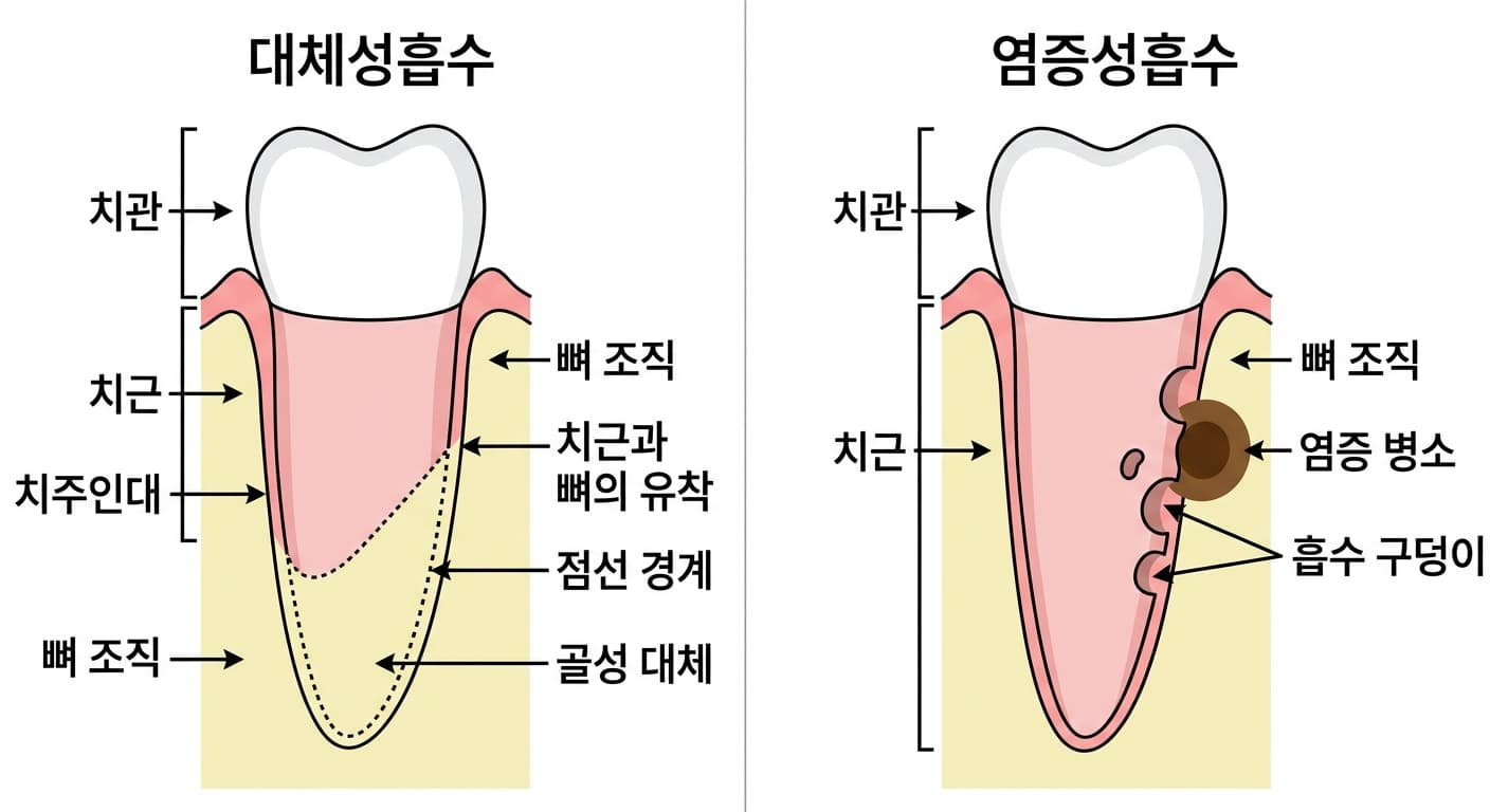
There are also complications caused by the trauma itself. When a tooth is completely avulsed and then replanted, if the periodontal ligament cells have dried out for too long or the damage is extensive, the root surface can end up in direct contact with bone without a periodontal ligament between them, and the root gradually turns into bone tissue through a process called replacement resorption. Conversely, if infectious material inside the root canal spreads into the periodontal tissue through the dentinal tubules, the root surface can develop pitted, uneven areas through a process called inflammatory resorption. Because these two types of resorption differ in cause and progression, they are managed differently.
Once replacement resorption begins, the tooth often gradually turns into bone and is eventually lost, so the prognosis is poor. Inflammatory resorption, on the other hand, can be halted by removing the underlying infection with root canal treatment and filling the canal with calcium hydroxide to calm the inflammatory response.
Course and Follow-up Care
Regardless of which treatment was performed, regular radiographic checkups every few months are needed to confirm that the periapical lesion is shrinking and that the root is growing as expected. In teeth that have undergone regenerative endodontic treatment, the canal has been reported to gradually calcify and narrow over time, with some teeth regaining a positive response to electric pulp testing, but not every tooth recovers to the same degree.
Even after the apex has closed and the lesion has resolved, if the tooth has discolored or the canal has become severely calcified, additional treatment such as root canal therapy or whitening may be needed later on. Above all, since a lack of symptoms right after a child's dental trauma cannot be taken as reassurance, it is important to have the tooth's progress checked at the dentist at set intervals after the injury.