
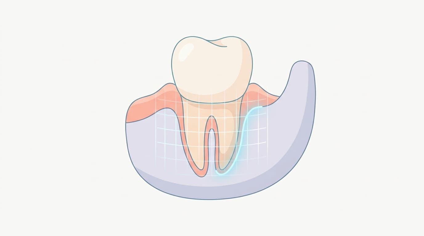
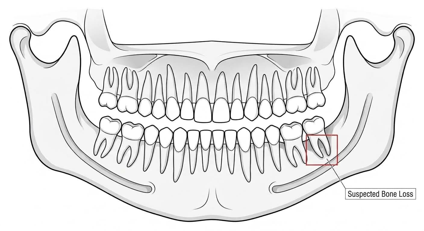
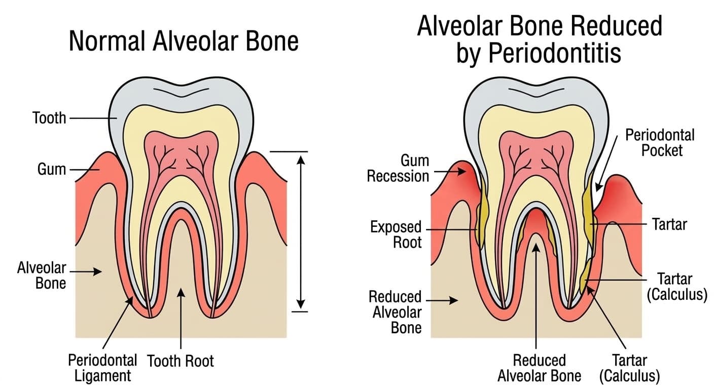
More dental clinics are having AI scan an X-ray first and mark how much the gum bone has receded. Alveolar bone is the jawbone that holds the teeth in place, and as periodontitis progresses, this bone gradually erodes and recedes. In a piece on AI that flags spots that look like cavities, a fair number of the flagged spots turned out not to be actual cavities. AI that detects bone loss is a somewhat different story. A recent review pooling multiple studies finds that its ability to spot areas of bone loss is fairly stable, but results vary depending on the image type and diagnostic task, and it is still too early to treat that output alone as a diagnosis of periodontitis.
Out of 100 X-Rays, How Many Does It Get Right?
A comprehensive 2025 review re-examined 30 studies on the topic, weighing their quality along the way. Pooling the ten studies whose data could be combined statistically, when shown 100 X-rays that actually had bone loss, AI correctly identified it in 87 of them, but out of 100 normal X-rays, it mislabeled 24 as showing gum disease. When the papers were graded using criteria built specifically for evaluating AI research meant for clinical decision-making, however, none scored at the highest confidence level, and 63 percent landed only in the middle tier. The performance numbers themselves aren't bad, but the studies that produced them are still uneven in design.
Panoramic vs. Per-Tooth X-Rays: Each Is Good at Something Different
Another meta-analysis published around the same time compared binary detection (disease present or not) against staging (mild, moderate, or severe). For binary detection, periapical X-rays (close-up shots of a single tooth) performed consistently well, with 87 percent sensitivity and 82 percent specificity. For staging, however, panoramic X-rays that capture the whole mouth in one image did better, with 89 percent accuracy. The researchers concluded that panoramic X-rays suit screening and staging, while periapical X-rays suit early, binary detection.
Two Tasks It Handles Especially Well
Narrowing the task pushes accuracy even higher. An analysis pooling eight studies and roughly 12,000 molar images that looked for furcation involvement (bone loss reaching the area between the roots of multi-rooted molars) recorded 93 percent sensitivity and 94 percent specificity, rising to 96 and 97 percent when limited to lower-jaw molars. Even so, the study was clear that a hands-on probing exam remains the reliable, essential test, and that X-rays and AI readings are only supplementary data.
An analysis pooling five studies and about 12,000 images looking for signs of bone loss around dental implants also found AI performing well, with 88 percent sensitivity and 91 percent specificity. Here too, the researchers drew a clear line: the result is only a reference point suggesting peri-implantitis, not a substitute for a clinical exam of the gum tissue itself.
Where Trust Is Still Lacking
By contrast, attempts to diagnose gingivitis or periodontal disease itself from images alone still produce inconsistent results. Studies diagnosing gingivitis from intraoral photographs stayed in a 74 to 78 percent accuracy range, while studies diagnosing periodontal disease itself from images varied widely, from 47 to 81 percent. Another comprehensive review from 2023 likewise concluded that, given the low quality of evidence accumulated so far and the wide performance gap between different programs, AI readings should be treated with caution rather than accepted outright as a diagnosis.
Worth Asking About in the Clinic
If you're told about an AI reading result, it's worth asking:
- Whether the image AI analyzed was a full-mouth panoramic X-ray or a close-up of an individual tooth
- Whether the flagged bone loss was also confirmed with a hands-on probing exam of the gums
- If it's around an implant, whether that area was separately re-examined
Bone height on an X-ray is just one of several criteria used to determine periodontal disease. How well the gum tissue is still attached and whether a tooth is loose can't be seen from an image alone; those are things a dentist still has to check in person. The best way to use this technology is to let an AI-flagged spot be a reason to get checked sooner, not later.