
When the Canal Cannot Be Found, Root Canal Treatment Stalls
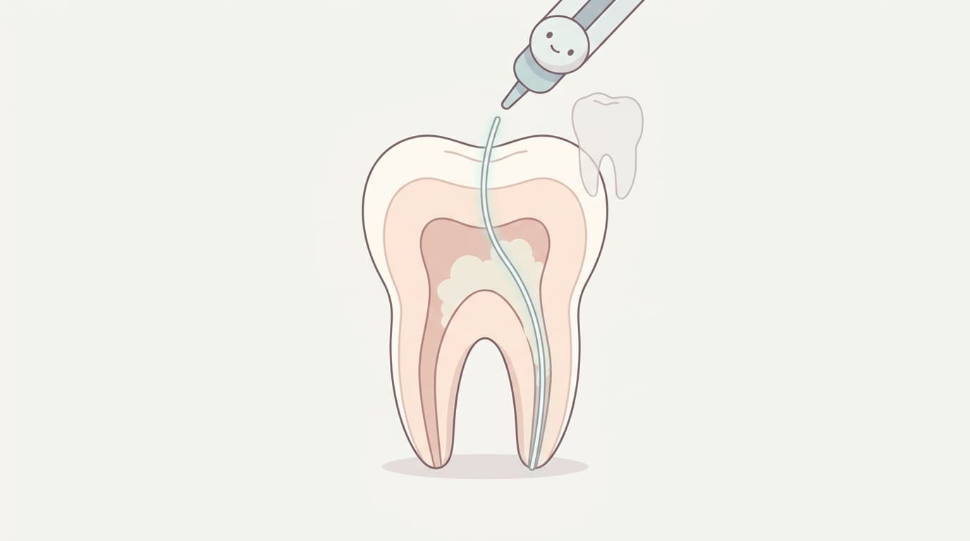
Root canal treatment (endodontic therapy) locates the canal inside a tooth, removes the infected tissue, and fills the space. But the canal inside a tooth can undergo calcific degeneration (pulp chamber calcification), in which calcified material narrows or blocks it. This is common enough to be observed in more than 60% of adult teeth, and it is known to appear more often in women than in men.
In a tooth that has suffered trauma, the canal narrows as excess dentin forms while the pulp tries to survive. With age, calcification gradually progresses throughout the entire pulp. In both cases, the canal's shadow on radiographs grows faint or disappears, making it hard for the clinician to locate the canal orifice with the naked eye or at low magnification. The more affected the tooth, the greater the risk of missing the canal entirely or drilling in the wrong direction and perforating the side of the tooth.
To solve this problem, dentistry has long relied on magnification equipment, and more recently, computer-based technologies have emerged that map out the path before surgery or indicate the location in real time during the procedure.
The Standard That Held: Microscopes and Ultrasonic Instruments
Until now, the standard way to locate a calcified canal has been to combine a dental operating microscope with ultrasonic instruments. The dental microscope magnifies the surgical field 2.5 to 30 times while illuminating it brightly; in the narrowed pulp chambers of elderly patients, magnification of 25x or higher is used to distinguish differences in shade, texture, and translucency between calcified deposits and the original pulp chamber wall. The diamond-coated tip of an ultrasonic instrument then removes the deposits little by little, allowing the clinician to reach the canal orifice without perforating the tooth.
This combination also plays a central role in microsurgical apicoectomy, in which the root tip is surgically exposed and treated. Conceptualized by Professor Syngcuk Kim in the early 1990s, microsurgical apicoectomy is performed at 8-10x magnification, with the resected root surface examined at a higher magnification of 15-25x. This is reported to have raised the success rate of traditional apicoectomy, which had remained at 44.2-60%, to as high as 91.1%.
Still, the microscope and ultrasonic instruments are, in the end, tools that the clinician must see and operate by hand. The direction of a canal hidden inside a tooth can only be predicted before surgery; magnification alone cannot reveal the entire path in advance.
3D Guides That Plan the Path Before Surgery
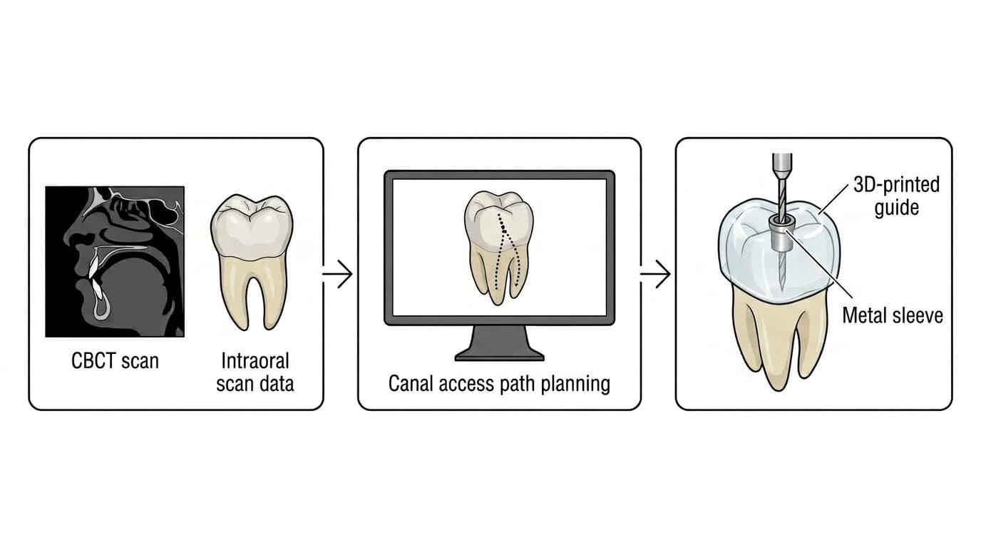
Static guides (guided endodontics) emerged to address this limitation. A computer overlays a 3D image of the tooth and jaw taken by CBCT with tooth surface data obtained from an intraoral scanner, allowing the access path to the canal to be planned before surgery. A 3D printer then fabricates a guide (stent) based on this plan, which is seated on the tooth; guiding a bur through the metal sleeve fitted into the guide lets the clinician reach the calcified canal orifice without unnecessary loss of tooth structure or risk of perforation.
A study by Buniag and colleagues reported a 1-year success rate of 91.7% for canal access using static guides, with particularly strong outcomes in difficult cases such as molars, the maxillary palatal root, and the mandibular distal root.
However, because a guide simply follows the path decided before surgery, the path cannot be changed during the procedure. Errors can also occur during guide fabrication, and if there is not enough room to open the mouth (interocclusal space), the guide itself may be difficult to seat.
Real-Time Navigation That Can Change Course Mid-Surgery
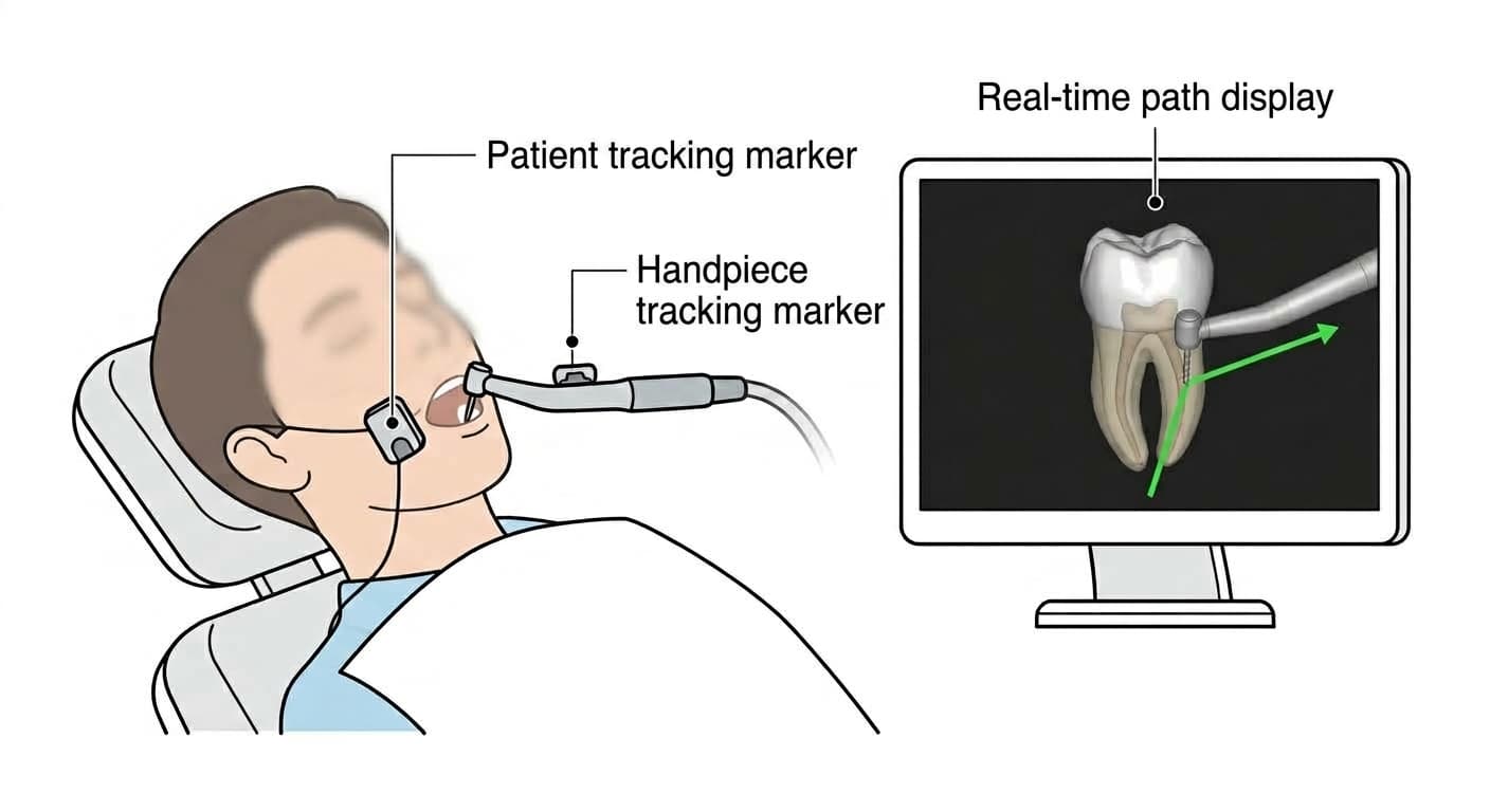
Dynamic navigation systems (DNS) go beyond the limitations of static guides. Infrared optical tracking markers are attached to the patient and to the dental handpiece, and the bur's position, direction, and depth are overlaid on the CBCT image in real time. Watching the screen, the clinician can adjust the path and angle on the spot, so the procedure does not block the operator's view the way a guide does, while still staying close to the planned path.
A study by Chen and colleagues found a CBCT-based 1-year success rate of 94.3% for dynamic navigation systems. Beyond accessing calcified canals, they are also used in demanding procedures that require both visibility and flexibility, such as removing separated instruments, apical surgery, and complex retreatment. More recently, the technology has been combined with ultrasonic retrograde filling and augmented reality (AR) display to improve both precision and procedure time.
Added Precision from Robotics, and What Remains
The most recent approach is robot-assisted surgery, which applies a haptic-feedback robot to microsurgical apicoectomy. The goal is for the robot to support the clinician's hand movements and reduce the variability in skill between operators. It has been shown to produce smaller angular and linear deviations and improved precision than static guides or dynamic navigation systems, and across guided surgical systems overall, including static guides, dynamic navigation systems, and robot-assisted surgery, 1-year success rates above 90% have been reported.
However, robotic systems are still expensive, and how practical they are in an actual clinical setting is still being validated. The technology for locating canals has advanced in precision, moving from microscopes to 3D guides, real-time navigation, and robotic assistance, but not every root canal treatment requires this equipment. In practices equipped with CBCT, an intraoral scanner, and guide-fabrication facilities, these technologies are used selectively for difficult cases such as severely calcified canals, missed canals, or failed retreatment.