
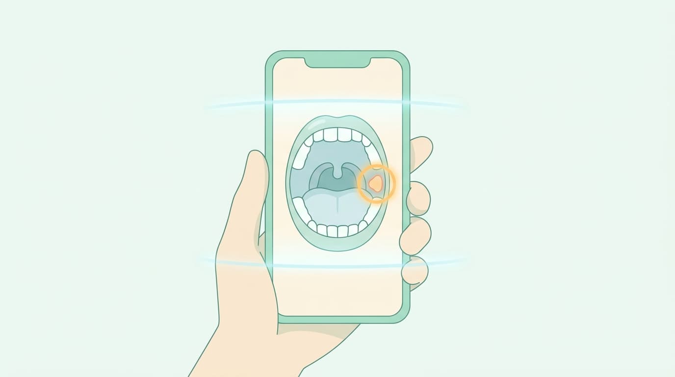
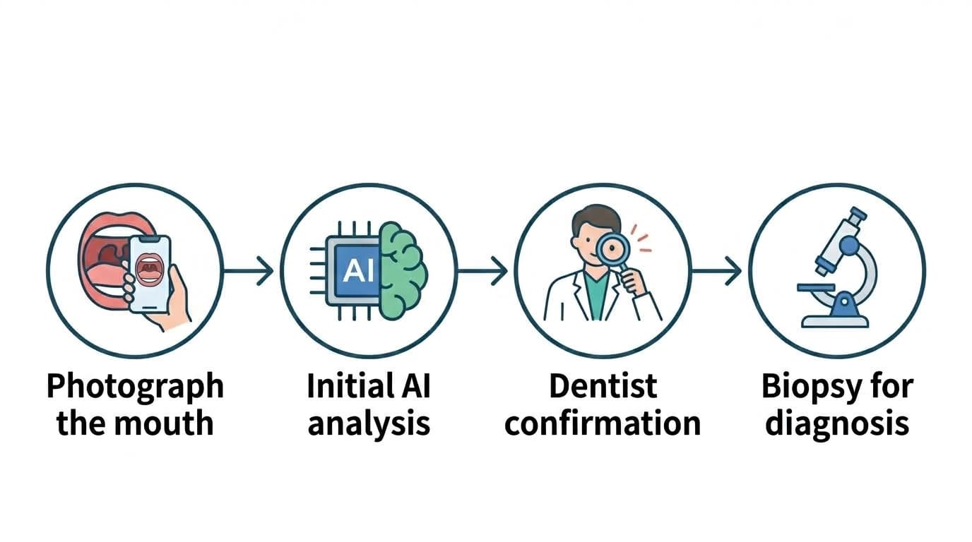
Services are emerging that let you photograph the inside of your mouth with a smartphone so AI can flag lesions that could progress to cancer. A 2026 meta-analysis found that this approach catches more than 80% of real lesions without missing them, but it also mislabels normal tissue as a lesion so often that it's difficult to use as a standalone diagnostic tool. Where AI Caries Diagnosis covered how accurately AI finds cavities, this article looks at whether a single photo can screen for oral cancer and its precancerous lesions, conditions with far heavier consequences.
What the Photos Are Trying to Detect
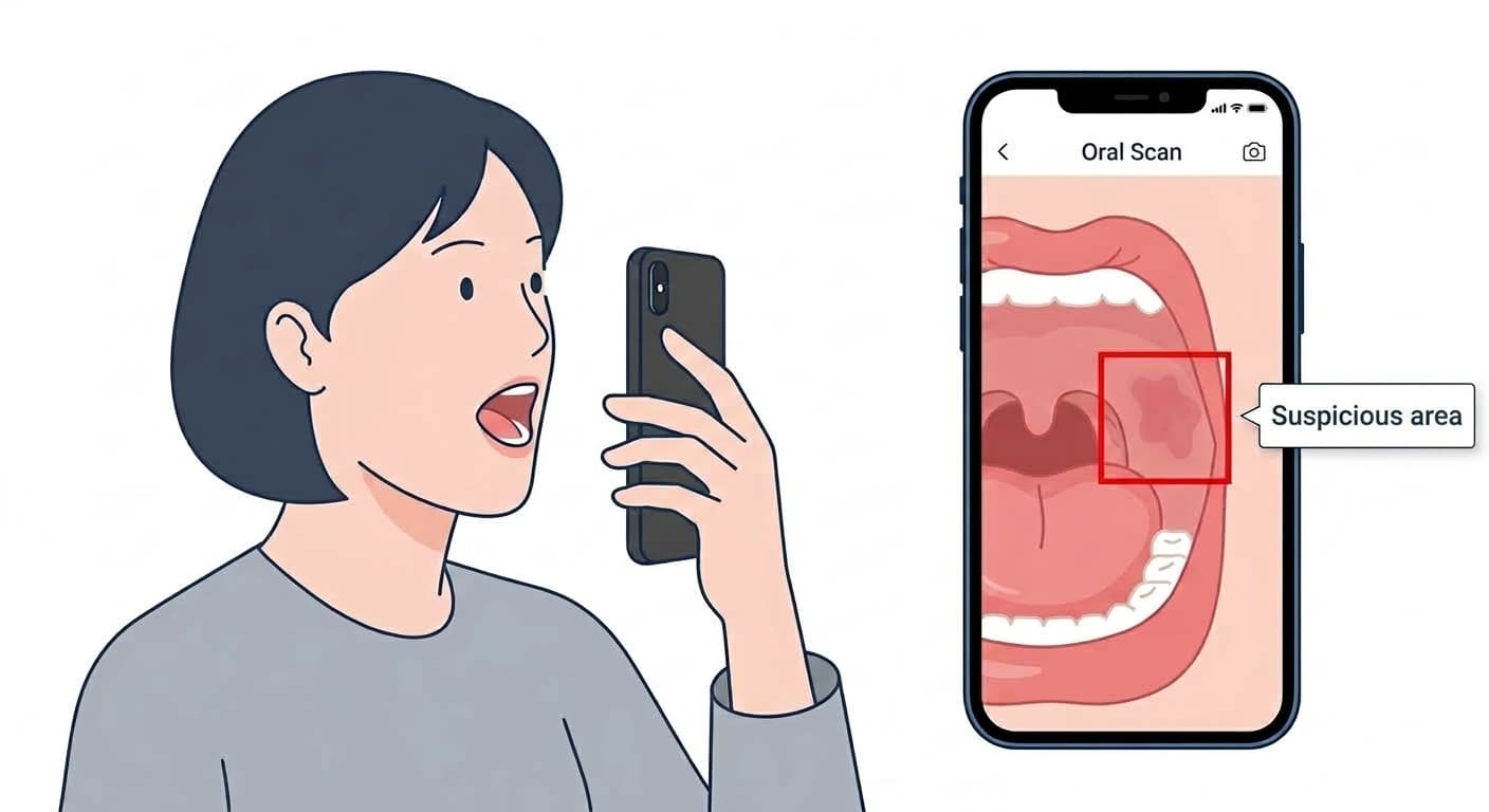
Oral squamous cell carcinoma, which accounts for about 90% of oral cancers, sees its five-year survival rate drop by nearly half the later it is found. Lesions that can progress to cancer, such as leukoplakia (white patches), erythroplakia (red patches), and oral lichen planus, often appear first, which is why catching them at this stage matters. AI models learn the differences in color, border, and texture between normal mucosa and lesions in mouth photos taken with a smartphone or digital camera, then either draw an outline around the suspicious spot or classify it as a lesion or not. Smoking, alcohol use, HPV infection, UV exposure, and chronic irritation or inflammation are cited as risk factors that raise the risk of these lesions and of cancer.
Special Light and Dyes Used to Be the Standard
Until now, the adjunctive tests dentists used to screen for suspicious oral cancer sites were autofluorescence devices, which shine light of a specific wavelength to make lesions stand out, and vital staining tests, which apply a dye to distinguish lesions. But the American Dental Association's latest clinical guideline, published in 2026, recommends against using these light-based adjuncts for routine screening, because the certainty of the evidence behind them was very low. It did make clear, however, that all adults, including those with no abnormal findings, should still get a visual and tactile oral examination. A review that pooled multiple non-invasive tests found that optical coherence tomography, spectroscopy that examines tissue with specific wavelengths, and brush cytology, which scrapes off cells for testing, actually showed the highest accuracy. Still, the guideline noted a limitation: many of the studies it drew on analyzed oral cancer and its precancerous lesions together rather than separately, and it left in place the fact that biopsy (removing tissue and examining it under a microscope) remains the diagnostic gold standard.
How Accurate Is Photo Analysis?
A 2026 meta-analysis pooled 21 relevant studies and statistically combined results from 12 of them with comparable conditions. Sensitivity for detecting lesions that could progress to cancer was 81%, and 84% for oral cancer itself, meaning relatively few real lesions were missed. The problem is specificity. It came in at just 16% for lesions and 14% for cancer, leading the researchers to state that using this approach alone would produce a very high rate of false positives. That means 84 to 86 out of every 100 normal sites could be flagged as suspicious. Even though specificity reported by individual studies ranged much higher, from 73% to 97%, the pooled statistical value dropped this sharply because photography methods, diagnostic criteria, and the target lesions varied enormously from study to study.
What Drives the Variation: Shooting Conditions
Even with the same AI, performance varied greatly depending on whether the whole mouth was photographed or just a cropped shot of the lesion, and camera type, lighting, and resolution also changed the results. More than half of the 21 studies reviewed (11 of them) were rated as having a high risk of bias, and only about half confirmed their findings with pathology testing. That means the rest measured AI accuracy against expert visual judgment as the reference standard, which could differ from real-world clinical performance where biopsies are actually done. The devices used for photography were a mix of smartphones and professional cameras, and there is still no common guideline for how the photos should be taken.
How to Read This in the Clinic
If an AI or a special-light device flags a suspicious spot, there's no need to immediately worry about cancer; the current evidence shows that mislabeling normal tissue as a lesion is common. On the other hand, no flag doesn't mean you can relax either. If you've smoked or drunk alcohol for a long time, or have a white or red patch in your mouth that won't heal, you should get a direct examination at a dental or oral and maxillofacial surgery clinic and, if needed, confirm it with a biopsy, regardless of what these adjunctive tests show. Adjunctive tests are there to support a dentist's visual examination, not to replace it.