
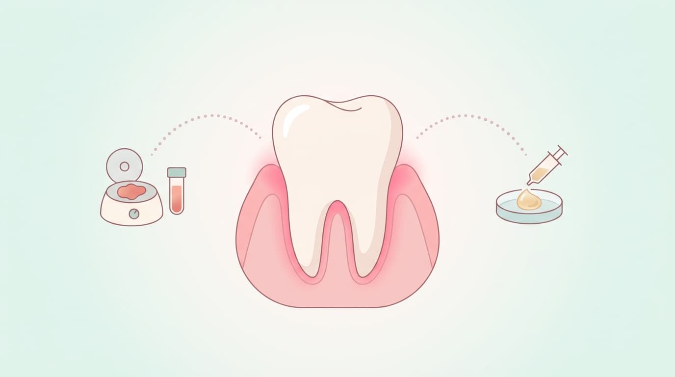
In regenerative surgery for localized periodontal defects (sites where alveolar bone has been lost), long-term follow-up results have been published comparing PRF (platelet-rich fibrin), made by centrifuging the patient's own blood, with Emdogain, an enamel matrix derivative gel. In two randomized controlled trials involving the periodontics research team at Semmelweis University in Hungary, improvements in periodontal pocket depth and clinical attachment level were maintained at 3 and 5 years. There was no statistically significant difference between the two materials in the main clinical indicators, but because these are small studies, this is not grounds to conclude that the two materials are equally effective.
Why Bone Loss From Gum Disease Doesn't Heal on Its Own
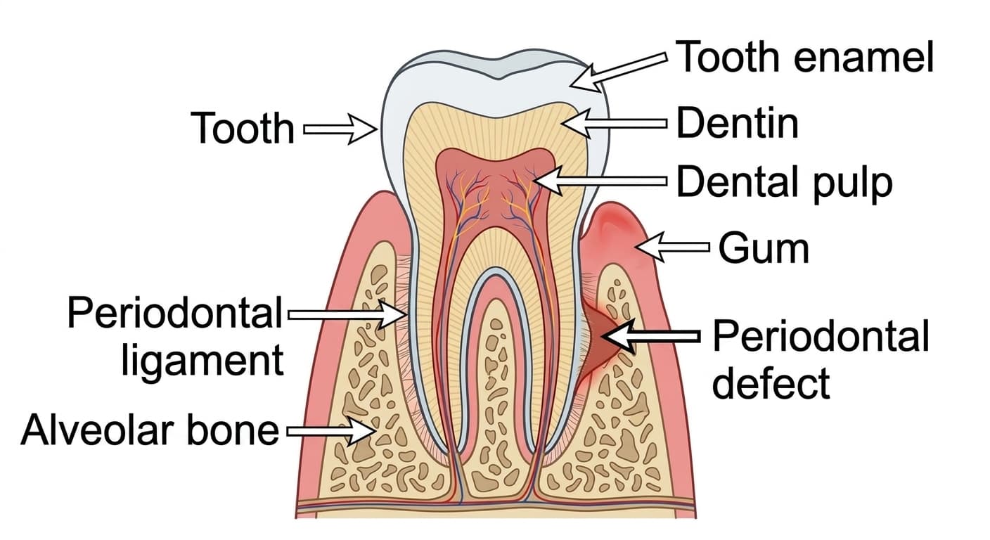
When periodontitis is left untreated for a long time, inflammation caused by bacteria destroys the periodontal ligament surrounding the tooth and the alveolar bone (the gum bone that supports the tooth). Treatment that simply calms the inflammation itself, the kind you can check with a periodontitis self-test kit, does not make bone that has already dissolved away grow back on its own. Refilling this site requires periodontal regenerative surgery, in which the gum is opened, the inflamed tissue is removed, and a material that induces regeneration is placed inside.
The material most widely used for this purpose over the years has been Emdogain. It is a protein mixture extracted from the enamel matrix of developing porcine tooth buds, with amelogenin as its main component. It mimics the signals of tooth development to induce regeneration of the cementum covering the tooth root, the periodontal ligament, and the alveolar bone.
A Regenerative Material Made by Spinning the Patient's Own Blood
PRF works on a different principle. When venous blood is drawn from the patient without any anticoagulant and spun directly in a centrifuge, it forms a clot in which platelets and white blood cells are trapped inside a dense fibrin mesh. When this clot is packed into the defect site, the various growth factors released from the platelets and white blood cells slowly leach out over about a week, helping new blood vessels grow and cells gather at the site.
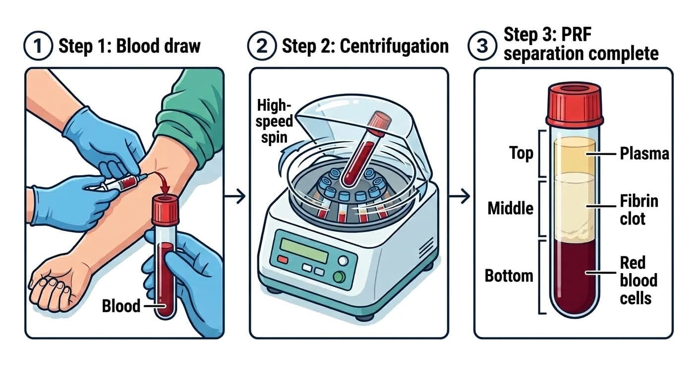
Since the original membrane form (L-PRF) was introduced, several variants have followed: A-PRF, which lowers the centrifuge speed to retain more cells, and i-PRF, a liquid form that can be injected before it clots. Unlike Emdogain, which must be purchased as a protein extracted from animals, PRF is drawn directly from the patient's own blood on the day of treatment, so material costs are lower and there is less concern about foreign-body reactions.
How Far Has Combination With Bone Graft Material Been Confirmed?
The effect of adding PRF to open flap debridement (surgery that opens the gum to clean the root surface and inflamed tissue) has also been studied. In a meta-analysis pooling 23 randomized controlled trials, the 16 trials that reported outcomes at 9 months found that the group given PRF showed an average 1.11mm greater reduction in periodontal pocket depth and a 1.13mm greater gain in clinical attachment level (an indicator of how much tissue remains supporting the tooth) than the group that underwent surgery alone. However, the certainty of evidence ranged only from very low to moderate, and results varied considerably between studies.
A network meta-analysis covering 32 studies found that combining PRF with bone graft material ranked highest for both reducing periodontal pocket depth and increasing bone volume. However, this ranking alone does not mean the combination is necessarily superior to PRF alone, or that it should be used for every defect; the evidence for some combinations is limited and needs further verification. In India, one study compared a combination that polymerizes injectable PRF together with Bontree, an octacalcium phosphate (OCP) bone graft material, against the bone graft material alone. Over 6 months of follow-up in 12 patients, the combined group showed a favorable trend, but the difference between groups was not statistically significant.
Clinical Improvements Maintained at 3- and 5-Year Follow-up
Studies have also directly compared PRF and Emdogain by following the same patients over a long period. In a clinical trial of 30 patients with stage 3-4 periodontitis, the PRF-treated group showed an average improvement of 4.40mm in periodontal pocket depth and 3.87mm in clinical attachment level six months after surgery, compared with 3.53mm and 2.13mm in the Emdogain group. Even after 3 years, these improvements remained largely unchanged in both groups, and no statistically significant difference between the groups was found in either periodontal pocket depth or clinical attachment level. Only gum recession was lower in the PRF group.
Another study compared the newer-generation A-PRF+ with Emdogain, starting with 30 defects in 18 patients and evaluating 26 defects in 14 patients at the 5-year mark. Improvements in clinical attachment level were maintained in both groups, with no statistically significant difference between them. In the 3-year study, 26 of the 30 patients initially enrolled completed follow-up. Both studies were small, and some researchers overlapped between them, so confirmation by other research teams is still needed.
Research so far supports the possibility of using PRF in periodontal regenerative surgery. Combining it with bone graft material ranked high on some indicators, and improvements in clinical indicators were maintained in long-term comparisons with Emdogain. However, larger-scale comparisons are needed to establish which material is superior or whether they are truly equivalent. Future research should distinguish between defect shapes and PRF preparation conditions to determine which combinations retain their benefit.