
White spots that spread across a tooth's surface, or early cavities hiding between teeth, no longer have to be drilled out. Resin infiltration (brand name Icon), which flows a low-viscosity resin into the lesion to seal it, has taken that role. But the treatment carried one weakness: treated spots still looked dark like cavities on X-rays, making it hard to tell from imaging alone whether the lesion had stopped or kept progressing. Researchers at Queen Mary University of London recently added a fluoride-releasing glass component to create a resin that shows up more clearly than tooth enamel on X-rays, and confirmed its performance in the lab.
How to Stop Cavities Without Drilling
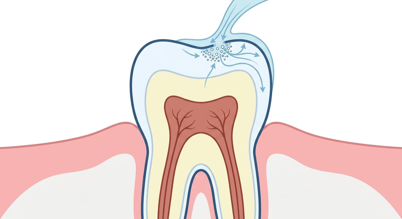
Resin infiltration is used for lesions that haven't yet broken through the surface, cases where the outside of the tooth looks intact, but minerals have leached out of the enamel underneath, leaving a dense network of tiny pores. Typical targets are white spot lesions left behind after orthodontic brackets come off, and early proximal cavities hidden between teeth that are hard to spot by eye. Where an earlier article covered attempts to reverse white spot lesions with remineralizing agents, this study addresses a technical weakness in the resin-sealing treatment itself. The procedure etches the surface lightly with acid to open an entry point, flows resin into the pores, then cures it with light. Once the resin fills the pores, the way light reflects off the tooth changes, so the white-looking spot returns to a translucency similar to the surrounding tooth, and the channels that let in bacteria and acid are sealed off. In a 7-year clinical trial run by a research team at Charité - Universitätsmedizin Berlin in Germany, only 9% of treated lesions progressed, compared with 45% of lesions that were simply monitored, cutting the risk of progression by 80%. An earlier systematic review reached the same conclusion: resin infiltration suppresses the progression of early caries.
X-Rays Couldn't Tell a Treated Spot From a Lingering Cavity
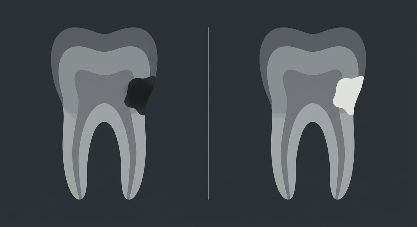
The problem showed up during follow-up. Cavities look dark on X-rays in proportion to how much mineral has leached out, meaning radiation passes through more easily. Even after resin infiltration fills those pores, the resin used until now blocks less X-ray radiation than tooth mineral, so it left a shadow on film similar to before treatment. The research team cited this as the motivation behind the new material: routine X-rays alone made it hard to distinguish a spot where the resin had stably filled the lesion from one where the cavity was still progressing. This weakness matters most for proximal lesions, which are hard to see directly and rely heavily on X-rays for follow-up.
A Resin That Shows Up More Clearly Than Enamel
The researchers created a new resin by mixing 30% fluoride-containing bioactive glass powder into the existing resin (Icon). This glass is a material already used in other dental fillings; it dissolves gradually in saliva, releasing calcium and fluoride ions. When the new resin was flowed into an artificially created early-caries model and examined with X-rays, it blocked radiation to a degree equivalent to 3.2 millimeters of aluminum, showing up more clearly than the comparison tooth enamel (2.0 millimeters). The degree of cure under light (conversion rate) was also higher, at 84.9% versus 74% for the existing Icon resin, meaning the resin itself hardened more solidly. After 23 weeks submerged in an acidic environment, it released more calcium and fluoride ions than in a neutral environment, and electron microscopy and X-ray diffraction analysis confirmed that new hydroxyapatite crystals (the same mineral found in tooth structure) had formed on the surface. In other words, in the acidic conditions where cavities tend to progress, the resin instead releases ions more actively and re-hardens the surface.
Still Just Results From a Tooth Model
These results come from an artificially created early-caries model in the lab, not from human teeth. Whether the same performance holds up in the mouth's environment, with saliva, bacteria, and chewing forces added in, and how clearly before-and-after treatment can be distinguished on an actual patient's X-ray, remain unconfirmed. Still, this study addresses, at the lab stage, one weakness that had long kept drill-free cavity-stopping treatment out of step with the follow-up imaging meant to track it.