
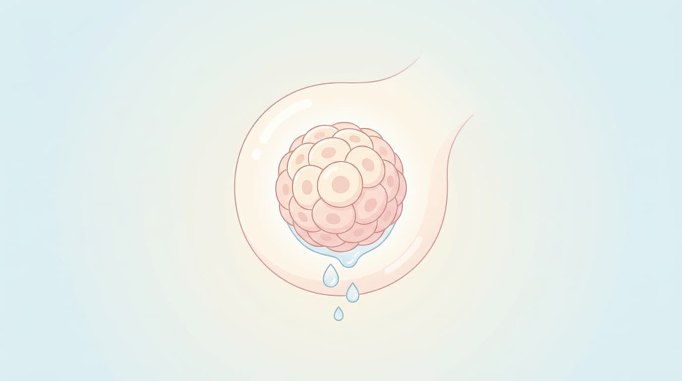
When Sjögren's syndrome or radiation therapy damages the salivary glands, treatment has mostly meant squeezing out whatever function remains from the surviving cells with medication, or getting by with artificial saliva. A study published in 2026 tried something different: growing entirely new gland tissue in the lab and transplanting it in. Human salivary gland organoids (small, three-dimensionally grown tissue masses built from stem cells), cultured without relying on any animal-derived components, were transplanted into Sjögren's syndrome mice, and their reduced saliva output increased.
Salivary glands grown without Matrigel behave like the real thing
To build a salivary gland organoid, stem cells taken from an adult salivary gland must be grown three-dimensionally in culture medium. Until now, this often relied on Matrigel, a gel extracted from mouse tumor cells, as a scaffold, but because its composition isn't precisely controlled, moving it into clinical use has been difficult. The research team grew organoids without Matrigel, using only a chemically defined culture medium combining the growth factors FGF10 and R-spondin1 with the antioxidant N-acetylcysteine. Removing even one of these three components sharply reduced the rate at which organoids formed.
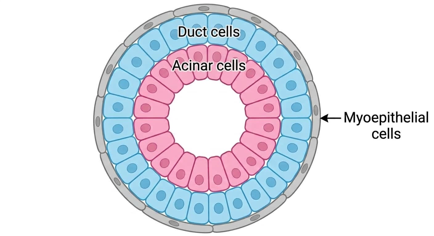
The organoids that grew this way had the same composition as a real salivary gland: acinar cells that produce saliva, duct cells through which saliva passes, and myoepithelial cells surrounding the duct. Single-cell genetic analysis also confirmed a differentiation pathway in which basal cells close to stem cells branched toward duct cells or acinar cells. When acetylcholine-class stimulants were introduced, intracellular calcium levels spiked instantaneously, the same response a real salivary gland shows when stimulated by the parasympathetic nervous system to secrete saliva.
Transplanted into Sjögren's syndrome mice, saliva flowed again
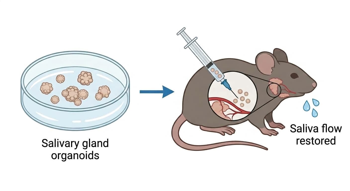
The research team transplanted these organoids directly into the salivary glands of mice used as a Sjögren's syndrome model. After 7 weeks, the amount of stimulated saliva was markedly higher than in mice injected with saline alone, and aquaporin-5, a water channel protein, also increased. Fluorescent tracking showed that the transplanted human cells had integrated into the mouse's existing duct structures, and 93 proteins found only in humans were detected in the saliva of the transplanted mice. This means the cells didn't just survive and settle in place, they actually produced saliva components.
An earlier study addressing salivary glands damaged by radiation aftereffects used an approach that inserted only a water channel gene into surviving duct cells. This study instead takes the approach of building an entirely new gland tissue and implanting it as a whole.
Gland structures also grew in the kidney, but there is no path to the mouth
The research team also implanted organoids beneath the surface of the kidney. Even in a location with no natural salivary gland environment whatsoever, mucus-secreting gland structures had formed by week 4, and by weeks 8 to 12 the tissue had grown into a more mature structure with blood vessels running through it. When early-stage mouse salivary gland connective tissue was implanted together with the organoids, the proportion of acinar cells was noticeably higher than with organoids alone, revealing that signals from surrounding tissue help drive maturation.
However, the tissue formed this way, even as it produced saliva, had no duct to carry it out into the mouth, so it simply pooled inside the tissue. Moving this into humans still requires a step connecting this reservoir structure to an actual salivary duct, and because all results so far come from mouse experiments, whether they carry over directly to humans must be sorted out in the next stage. Even so, this is the first time that, at the animal-experiment stage, building an entirely new salivary gland tissue rather than reviving dead salivary gland cells has been shown to restore saliva secretion.