

A joint research team from Lanzhou University and the Chinese Academy of Sciences in China has published a method for detecting abnormal temporomandibular joint (TMJ, the joint connecting the lower jawbone and the skull) motion using an earplug-shaped sensor, in the international journal ACS Sensors. Temporomandibular disorders are typically evaluated through symptom history-taking and clinical examination, with imaging added when necessary. The research team reported 91.7% accuracy in a preliminary classification of the signals collected from the sensor using an artificial intelligence model. This figure does not represent confirmed diagnostic performance in general patient care.
Measuring the Pressure That Presses on the Inner Ear Canal Wall
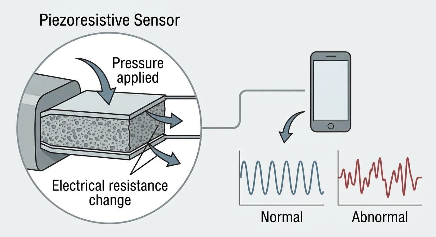
The temporomandibular joint sits right in front of the ear, against the anterior wall of the ear canal (the passage running from the ear opening to the eardrum). When the lower jawbone moves as a person opens their mouth or chews, that pressure is transmitted directly to the anterior ear canal wall as well. Using this principle, the research team processed a porous material made by reducing graphene oxide (an rGO aerogel) into a piezoresistive sensor and assembled it into an earplug-like form that fits into the ear. A piezoresistive sensor is a material whose electrical resistance changes according to the intensity of applied pressure; measuring this resistance change converts the magnitude and speed of the pressure into numerical values.
The sensor detected pressure changes as small as 14.5 pascals (Pa). Its response time was a fast 0.064 seconds, and its performance remained unchanged even after 1,000 repeated press-and-release cycles. It was sensitive enough to distinguish subtle facial movements such as swallowing or opening the mouth. The research team trained an artificial intelligence classification model called a support vector machine (SVM) on the signals collected this way, enabling it to distinguish the signal patterns of normal versus abnormal lower jaw movement.
Could It Replace Palpation and Imaging Tests?
The existing methods for checking temporomandibular disorders were palpation, in which a dentist presses on the joint area by hand to check for sounds or pain, and imaging tests using MRI or cone-beam CT (a three-dimensional X-ray scan that images the jawbone in three dimensions). Palpation relies on the examiner's tactile sense, so judgments can vary from person to person; cone-beam CT involves radiation exposure; and while MRI does not use ionizing radiation, its cost and examination time must be considered.
Attempts to capture TMJ motion mechanically are not new in themselves. Joint vibration analysis (JVA), which records the vibration sounds from the joint using a skin-mounted microphone, has been used since the 1990s, but a 2013 review that pooled 15 related studies concluded that the evidence supporting its diagnostic reliability was insufficient. This sensor takes a different approach, directly measuring the physical deformation of the ear canal wall rather than sound. The research team confirmed that different signal patterns appeared in people with normal lower jaw movement versus those with abnormalities. This result was also consistent with clinical assessments and bone structure findings observed on cone-beam CT.
Still at the Laboratory Stage
This is an early-stage study conducted on a small number of volunteers, and it is proposed not as a definitive diagnostic test but as a screening tool to first flag possible abnormalities. The 91.7% accuracy figure is also derived only from the participants in this study, so validation involving a larger population is needed.
TMJ abnormalities often appear together with bruxism, the habit of grinding one's teeth. Whereas the approach of using a vibrating splint to reduce bruxism duration is a treatment that directly addresses jaws and teeth that are already grinding, this technology is closer to diagnosis and screening: first identifying how the TMJ moves.
This study demonstrates the possibility of confirming TMJ motion with an objective numerical measurement simply by inserting a sensor into the ear, without palpation or imaging tests. Further validation is still needed before mass production or clinical application, but the study is meaningful in proposing a new measurement principle that directly measures pressure deformation instead of listening to vibration.