

A dental research team at UCLA in the United States has suggested a way to shorten the time it takes ultraviolet light used in implant surface treatment to break down organic matter. In an experiment published in 2023, exposing a quartz ampoule to vacuum ultraviolet light at a wavelength of 172nm decomposed more than 90% of methylene blue, a model organic compound, within just one minute.
Treating the Surface Rather Than Changing the Implant
Photofunctionalization is a treatment that uses light to change the properties of a titanium surface. A review compiled by the UCLA research team explains that ultraviolet treatment reduces hydrocarbons on the surface, increases hydrophilicity so that water spreads more readily, and also changes the surface's electrical properties. These changes are the starting point for research into cell attachment and proliferation, and into osseointegration, the process by which an implant bonds with bone.
While guided and robotic surgery addresses where an implant is placed, photofunctionalization addresses the conditions under which a material's surface meets cells. The results of these two technologies therefore cannot be compared using the same accuracy figures. To read the results of a surface treatment correctly, you first need to look at what was treated and what change was measured.
What Does the '90%' Figure Actually Measure?
The research team placed a methylene blue solution in quartz ampoules, with and without a titanium specimen inside, and compared four types of ultraviolet light. Vacuum ultraviolet light reached its maximum level of decomposition within one minute, while the conventional UVC light source used for comparison took 20 to 30 minutes. This time frame applies specifically to the dye-decomposition conditions used in this experiment.
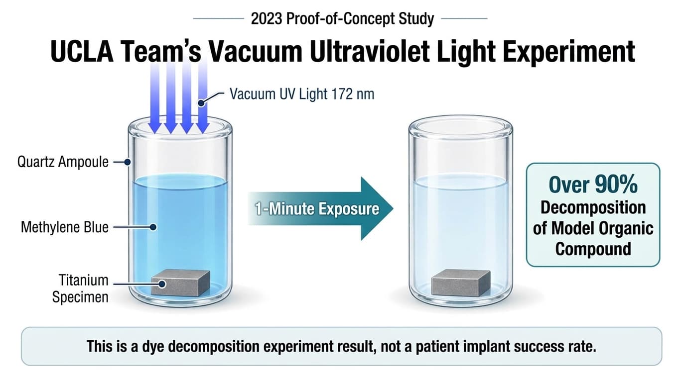
This is an explanatory diagram created based on the experimental conditions reported in the paper. It is not an actual photograph of the experiment.
When the quartz ampoule was replaced with a plastic tube, the decomposition effect dropped sharply. This means that achieving rapid treatment requires designing not just the wavelength of light but also the container and exposure conditions together. The figure of 'over 90%' likewise refers to the amount of dye decomposed under these specific conditions, and it cannot be read as the rate at which contamination is removed from an implant surface, or as a patient's treatment success rate.
Connecting Laboratory Speed to Clinical Outcomes
A follow-up review published online in 2024 expanded the scope of photofunctionalization research beyond osseointegration to include soft-tissue and bacterial responses. It examined how chemical changes on the surface lead to cellular responses, and it also covered studies involving patients. Even within the same field of research, material experiments and clinical studies answer different questions.
The vacuum ultraviolet experiment presented by the UCLA research team is an achievement in comparing light sources for surface treatment and specifying the conditions involved. To apply the result of rapidly decomposing a dye to actual patients, the treatment's effect on real implants, its impact on osseointegration, and long-term outcomes must be confirmed separately. Only by maintaining this distinction can the figure of 'one minute' accurately describe the development achievement it represents.