Overview
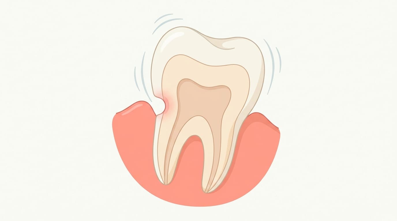
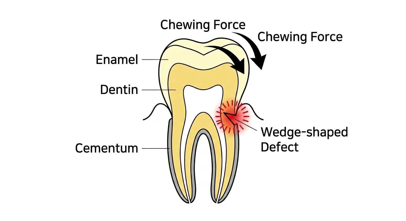
Abfraction is a condition in which abnormal chewing forces flex a tooth, concentrating tensile force (a pulling force) at the cervical area where the tooth meets the gum, and sharply wearing away the enamel and dentin into a wedge shape. Unlike a cavity, in which bacteria dissolve tooth structure, abfraction is a non-carious defect in which tooth structure fractures away microscopically under force. The defect has sharp, well-defined margins and can occur on a single tooth or appear on several teeth at once.
Causes
The most common cause is an occlusal factor that concentrates abnormal force on a tooth. When this force is repeated, the tooth flexes microscopically, and the concentrated force at the cervical area builds up fatigue cracks where the enamel and dentin meet. Abfraction is also known as a multifactorial condition that commonly appears alongside other causes. When abrasion from toothbrushing or chemical erosion from acidic foods or acid reflux overlaps with this process, loss of tooth structure progresses faster. Attrition, a separate form of wear that occurs on the chewing surfaces and incisal edges when teeth grind against each other, is often observed together with abfraction in the same patient.
Symptoms
A V-shaped or gently saucer-shaped defect can appear where the tooth meets the gum. The shape and margins of the defect vary depending on the cause and stage of progression, and an early lesion may not be very noticeable. If the defect deepens and exposes dentin, brief sharp sensitivity can occur with cold water, sweets, or cold air, through the same mechanism as dentin hypersensitivity. However, when the defect is shallow, it can also progress slowly without noticeable discomfort, so the absence of sensitivity does not mean the defect is absent.
Diagnosis
The dentist first examines the defect visually to check its shape and margins. However, the shape of the lesion alone makes it difficult to distinguish the cause. So the location, shape, depth, and surface condition of the lesion are recorded, and toothbrushing habits, intake of acidic foods, acid reflux, and habits such as bruxism or clenching are also checked to determine the cause comprehensively. Whether the defect is progressing and the degree of sensitivity are also considered when deciding on a treatment plan.
Treatment & Where to Go
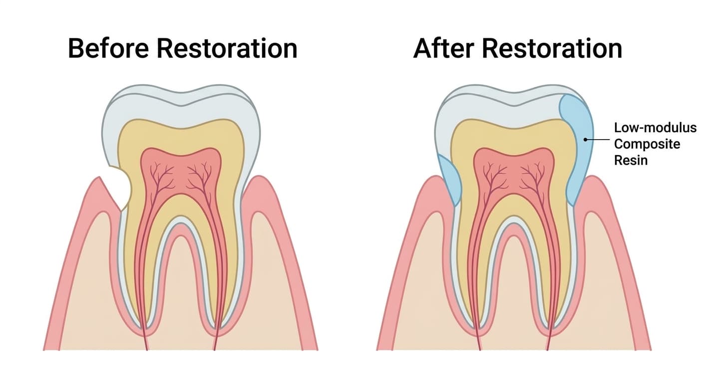
If the defect is shallow and there is no sensitivity or cosmetic concern, it can be monitored rather than filled right away. Restorative treatment is performed when there is a deep defect, sensitivity, a cosmetic concern, or a risk of gum recession or nerve exposure. Because a tooth flexes microscopically with every bite, filling with a flowable resin or glass ionomer material that has a low elastic modulus and can flex along with the tooth is recommended. If gum recession is also present, the location of the defect, the shape of the cervical area, and the condition of the exposed root are evaluated, and restorative treatment and periodontal plastic surgery may be performed alone or together. This care is provided by a dentist who performs tooth restoration, and coordination with a periodontist may be needed if gum problems are also present.
Outlook & Precautions
If the underlying force remains, the defect can continue to progress. Without correcting habits such as bruxism, clenching, or chewing predominantly on one side, the defect can recur in the same spot or on other teeth even after restorative treatment. If the defect feels like it is deepening or sensitivity worsens, it is advisable to see a dentist to check the depth of the defect and its distance from the nerve.