Question
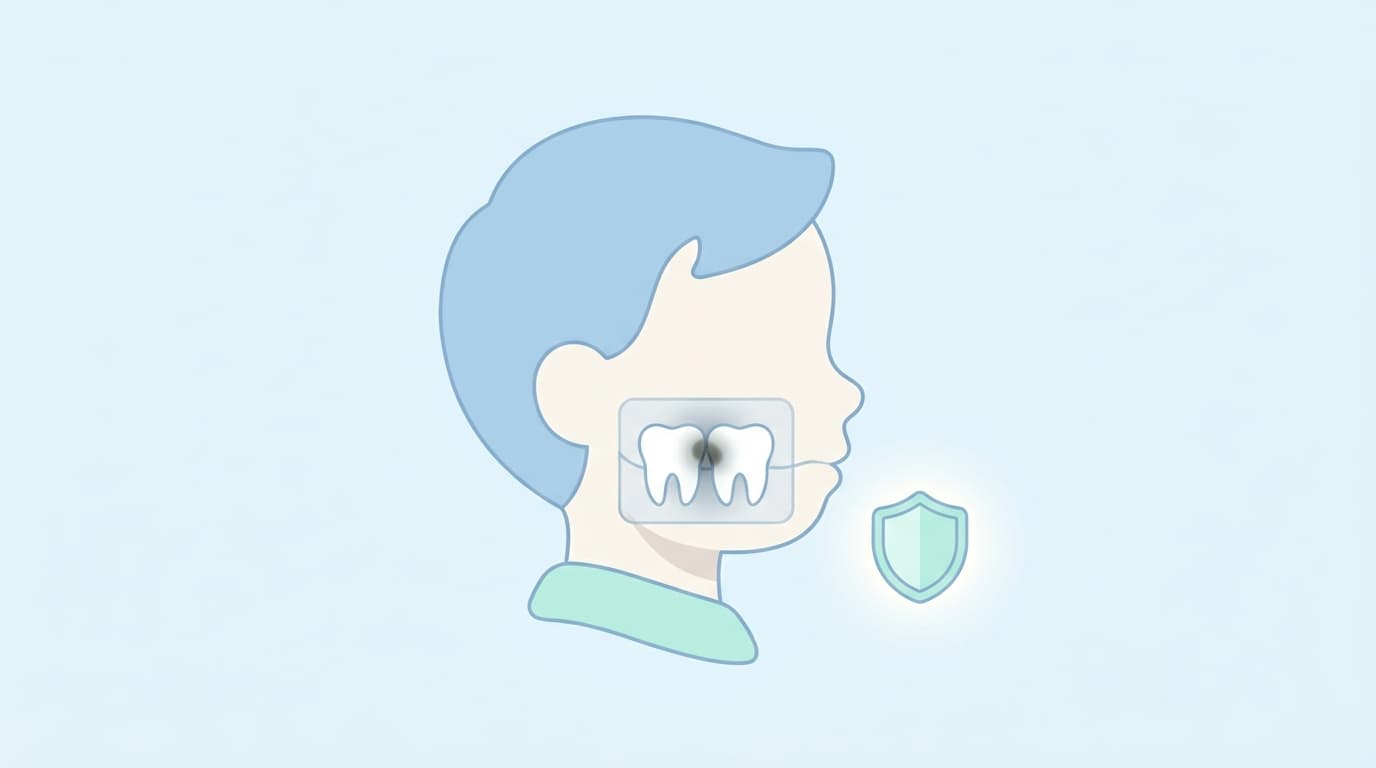
My six-year-old's dentist wants to take an X-ray at every visit. I'm worried about whether it's okay to take radiographs this often on a child who is still growing.
Short Answer
Dental X-rays for children are taken to diagnose areas that are hard to check by looking or by touch, such as cavities that have formed beneath the surfaces where two teeth touch. A child's tissues are more sensitive to radiation than an adult's. For that reason, the imaging itself is adjusted to keep exposure as low as possible while still obtaining the information needed for diagnosis.
There is no set number of X-rays that are considered safe, nor a fixed imaging schedule applied to every child alike. Whether an X-ray is needed at that point, along with the child's dental development stage and how prone they are to cavities, determines whether and how often imaging is done.
Why Is That?
X-rays Reveal What the Eye Can't See
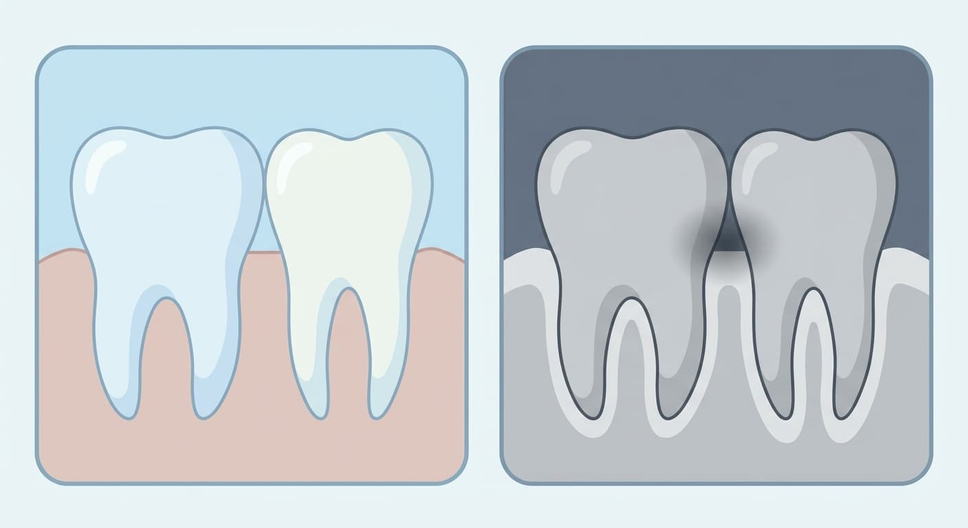
The surfaces where two teeth touch each other are hard to check by sight or with a dental probe. When tooth decay forms in this area, it often progresses on the inside without showing obvious signs on the surface. For that reason, a bitewing radiograph, taken by biting down on a film or sensor between the teeth, is used to reveal shadows beneath the boundary between enamel and dentin. How this exam detects interproximal caries has been explained in more detail in Why Bitewing Radiographs Are Used to Check for Cavities Between Teeth.
The same image can also show new decay beneath an existing filling and the height of the surrounding bone. Being able to check several things at once is one reason this exam is favored as a diagnostic tool.
Children Are More Sensitive to Radiation, So the Imaging Itself Is Adjusted to Lower Exposure
Radiation protection follows a principle that exposure should be kept as low as reasonably achievable while still meeting the diagnostic purpose. This is known as the ALARA (as low as reasonably achievable) principle. In children and adolescents, immature tissue that is still developing is especially sensitive to radiation, and the distance to the gonads (reproductive organs such as the ovaries or testes) is shorter, so the same dose of radiation can carry a greater risk than it would for an adult.
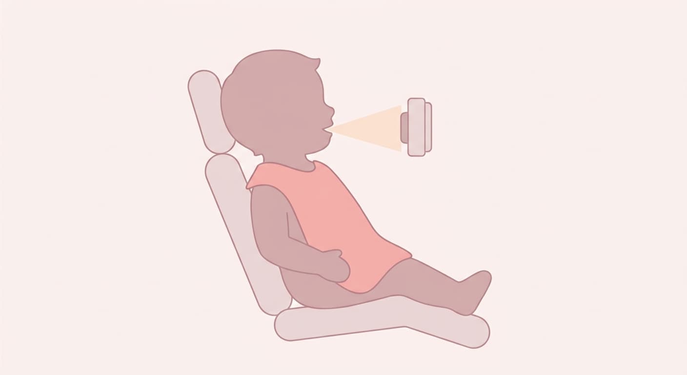
For this reason, several measures are applied together when imaging a child: a lead apron is used to block radiation from reaching areas outside the imaging site, the X-ray beam is narrowed to cover only the area that needs to be imaged, high-sensitivity film or digital sensors that produce a clear image with a lower radiation dose are used, and the equipment's exposure settings are adjusted so that only as much radiation as necessary is used. These measures don't work by reducing how often X-rays are taken, but by lowering the radiation dose used in each individual image when one is needed.
Whether and How Often to Image Is Decided Individually
When deciding whether to take an X-ray, the dentist considers the child's dental development stage (primary dentition, mixed dentition, permanent dentition, and so on), whether it is a first visit, and how prone the child is to cavities. Even at the same age, these factors can change when imaging is needed and how often.
How prone a child is to cavities, known as caries risk, varies from child to child. This risk is assessed by weighing risk factors, such as cavity-causing bacteria, frequent sugar intake, or reduced saliva flow, against protective factors, such as fluoride exposure or normal saliva flow. How this risk is assessed has also been covered in If a Cavity Doesn't Hurt, Is It Okay to Delay Treatment?. If the risk is judged to be high, the next imaging may be scheduled sooner so that new decay can be caught earlier; if the risk is low, a longer interval may be used for follow-up. Imaging is decided this way, tailored to each child's individual condition, rather than following a fixed number or schedule applied equally to every child.
Three-Dimensional Imaging (CBCT) Is Used Selectively in Other Situations
Most of the images taken at a child's routine checkups are two-dimensional, capturing the space between teeth or the whole tooth in a flat image. Cone-beam computed tomography (CBCT), by contrast, is equipment that produces a three-dimensional image and is used selectively only when needed, such as when the precise position of a tooth inside the jawbone, as with an impacted tooth, must be assessed. CBCT delivers a lower radiation dose than a conventional medical CT scan but a higher one than routine dental X-rays, so the same ALARA principle applies here as well, and it is taken only when judged truly necessary. Repeated imaging at routine checkups therefore does not add up to the level of exposure involved in a three-dimensional scan.