Question
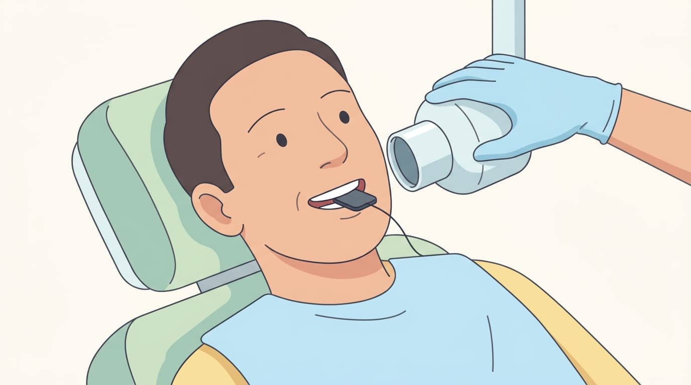
At every regular checkup, the dentist recommends a bitewing radiograph (where you bite down on a small film or sensor) to check the area between the teeth (the interproximal surfaces where teeth touch). The teeth don't look like they have cavities, and there's no sensitivity or pain, so I'm curious why this separate x-ray is needed and what it actually shows.
Short Answer
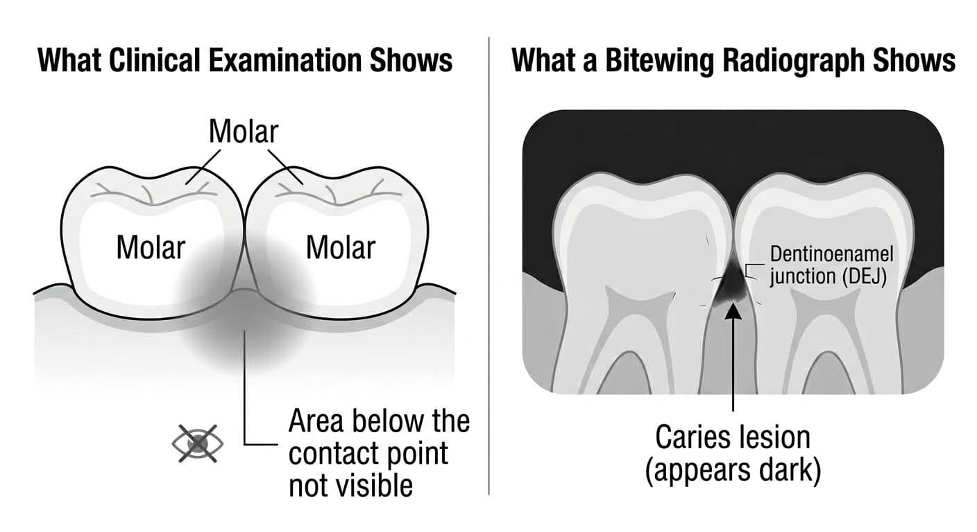
The area just below where two teeth touch is difficult to check by looking at it or feeling it with a probe. Cavities that start in this area often progress inward before the outer surface has even broken through, so they tend to show up on a radiograph, as a shadow (radiolucency), before they're visible to the naked eye. A bitewing radiograph is especially useful for checking this area because it captures both the interproximal surfaces between teeth and the area beneath the margins of existing fillings, making it well suited to detecting cavities here as well as recurrent ones. If something suspicious appears on the image, whether treatment is actually needed is decided together with what's seen during the clinical exam.
Why Is That?
Why Is the Area Between Teeth Hard to Check by Sight or Touch?
The area just below where two teeth touch is a spot where plaque (dental biofilm) easily builds up. Cavities that start here tend to spread wide at the outer surface and narrow into a cone shape as they move inward toward the dentinoenamel junction (DEJ, where enamel meets dentin). This means that even when there's no visible change on the outside, the cavity may already be progressing on the inside. Because the two teeth are pressed together, it's hard to see the inside of the contact area directly, and pressing hard with a probe tip to check it can actually be risky; the probe can break through an already-weakened enamel surface and create a new opening for bacteria to enter. For this reason, when checking this area, dentists use a blunt-tipped probe and only carefully feel for surface roughness. When a definitive diagnosis is really needed, an orthodontic separating band can be used to briefly open up the space between the teeth and view the contact area directly, but this isn't practical to do at every checkup.
What Does the Radiograph Show?
A bitewing radiograph is especially useful for observing new cavities on interproximal surfaces and recurrent cavities beneath the margins of existing fillings. Below the contact point, the inner enamel near the dentinoenamel junction lets more radiation pass through than normal tooth structure, so it appears dark on the image, and this dark shadow reveals the location and extent of early lesions. In a tooth that's already been filled, this also makes it possible to tell whether the area beneath the filling margin remains sound or whether a new cavity is spreading along the margin.
The lesions found can range widely, from shallow ones that don't even reach halfway through the enamel to ones that have progressed deep into the dentin, and how they're managed afterward depends on which stage they're at. Shallow lesions confined to the enamel are often not drilled right away; instead they're monitored while encouraging remineralization (the process of replacing lost minerals). Lesions that have crossed into the dentin, on the other hand, often lead to restorative treatment.
Why Take It Repeatedly, Not Just Once?
Studies that have tracked early enamel lesions that haven't yet cavitated show that a substantial share either stayed the same or actually remineralized, with only some progressing to an actual cavity. So finding an early lesion between the teeth doesn't automatically mean treatment is needed right away; what matters more is comparing over time whether the lesion is growing or staying still. People who are more prone to cavities have bitewing radiographs repeated every 3 to 6 months so this change isn't missed, and the results are used as an indicator for deciding whether restorative treatment is needed.
Why It's Needed Even in Teeth That Are Already Filled
Over time, a microscopic gap (microleakage) can form between a filling and the tooth, and if the original treatment didn't fully remove the cavity or the margin wasn't finished smoothly, a new cavity called secondary caries can start underneath. This area, too, is hidden from view by the filling, so a bitewing radiograph is used to check whether the area beneath the margin remains sound. If a new lesion is suspected beneath the margin, this leads to a decision to remove the existing filling and restore the tooth again.