
Question
I can see a dark spot on the chewing surface of my molar, but I don't feel any sensitivity or pain at all. Since it doesn't hurt, I'm wondering whether it's okay to just keep watching it instead of going to the dentist right away, or whether I should get it checked now.
Short Answer
Neither the absence of pain nor the visible dark spot, by itself, determines whether treatment can be delayed. What matters is whether the spot is a lesion that is still actively progressing or one that has already arrested, and whether the surface is still intact or has already broken through into a cavity. These three findings can only be judged by looking at and feeling the lesion directly, so it's hard to answer without examining it in person.
If the lesion has arrested, simply monitoring it without restorative treatment is often enough, unless there's an aesthetic concern. Conversely, if it's an active lesion that has already broken through into a cavity, progression tends to speed up from that point on and rarely stops on its own, so intervention is usually needed. Rather than "it doesn't hurt, so it can wait," the more accurate approach to this question is "decide only after the current state has been checked."
Why Is That?
Dark spots appear in both active and arrested lesions
Dental caries is a dynamic process in which acids produced by bacteria in the biofilm (dental plaque) that builds up on the tooth surface cause demineralization, the loss of minerals from the tooth, alternating with remineralization, the process by which minerals are redeposited. If this process is still tipped toward mineral loss, it's called an active lesion; if the loss has stopped and the site has settled into a more resistant state, it's called an arrested lesion.
What distinguishes the two states is not color but the texture of the surface and whether biofilm is attached. An active lesion is observed as a rough, soft surface covered with a thin layer of biofilm, where acid production continues. An arrested lesion has a hard, smooth surface that is easy to clean because no biofilm remains, and a crystal structure more resistant to acid (fluorapatite) has often formed there, making it even more resistant than the surrounding tooth structure. A spot that is more darkly stained black or brown is more likely to be a long-standing, arrested lesion, but color alone isn't conclusive; the surface must be felt directly to confirm.
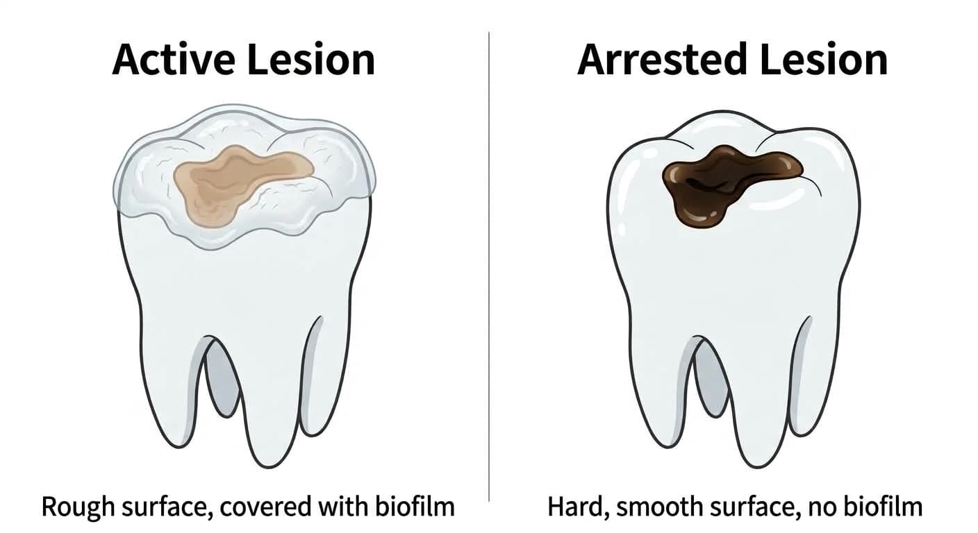
In the pits and fissures on the chewing surface, lesions are indeed observed to arrest over time, as chewing pressure flattens the grooves after the tooth erupts and self-cleaning improves. These arrested, inactive lesions often don't require invasive restorative treatment.
What matters more than the dark spot itself is how far it has progressed
An early lesion whose enamel surface hasn't yet broken through (such as a white spot lesion) doesn't necessarily progress to a cavity if left alone. In one longitudinal study conducted in a fluoridated water area, of 72 early white spot lesions, only 13% had progressed to a carious cavity after seven years, while more than half, 51%, had remineralized to the point of being indistinguishable from normal enamel. In other words, the mere presence of an early lesion doesn't necessarily mean it will only get worse.
There is, however, one clear turning point: the moment the enamel surface actually breaks through into a cavity. Past this point, a lesion very rarely stops on its own, progression speeds up, and it usually leads to restorative treatment. So the truly important question when looking at a dark spot isn't "how dark is it" but "is the surface still intact, or has it already broken through." Dentists assess this on a staged scale, from sound enamel all the way to a wide cavity that has spread into the dentin, and this stage determines whether to encourage remineralization or move straight to a restoration.
In areas that are hard to see from the outside, such as the inner slopes of the chewing surface or between teeth, visual inspection alone may not be enough to judge how far a lesion has progressed. In these cases, a bitewing radiograph, taken by biting down on a film between the teeth, is used to check whether the shadow has spread beneath the enamel and into the dentin.
Management also depends on the individual's caries risk
Even for lesions at the same stage, the intensity of management differs depending on the person's overall caries risk. Pathological and protective factors, such as whether active early lesions are present, whether restorative treatment was received within the past three years, and whether saliva flow is reduced, are weighed together to classify a person as low, moderate, or high risk. Those classified as high risk are typically recommended a tighter regimen: checkups every 3 months, application of a high-concentration fluoride coating (fluoride varnish) to promote remineralization at each visit, use of prescription-strength high-concentration fluoride toothpaste, and radiographs every 6 to 12 months. For someone at low risk, monitoring an arrested lesion of the same severity at longer intervals may be enough. So "is it okay to just watch it" is a question that can only be answered by looking at the lesion's condition together with the person's overall risk level.
How an active, progressing lesion is managed
If the lesion is active but the surface hasn't broken through yet, the first approach is often to try to halt and reverse progression through fluoride varnish application or induced remineralization, without restorative treatment. Fluoride application isn't exactly the same process as remineralization, and how fluoride application and remineralization differ has been explained separately. For an early lesion on a smooth tooth surface or an interproximal contact area, resin infiltration, which seals the lesion by infiltrating resin into its microscopic pores, is also used to stop progression without a restoration.
On the other hand, a lesion that has already broken through into a cavity and spread into the dentin is, in most cases, moved to restorative treatment, removing the damaged tissue and filling it, rather than waiting for it to stop on its own. The answer to "is it okay to delay" doesn't depend on pain, but on what stage the lesion is at now and what the person's risk level is.