
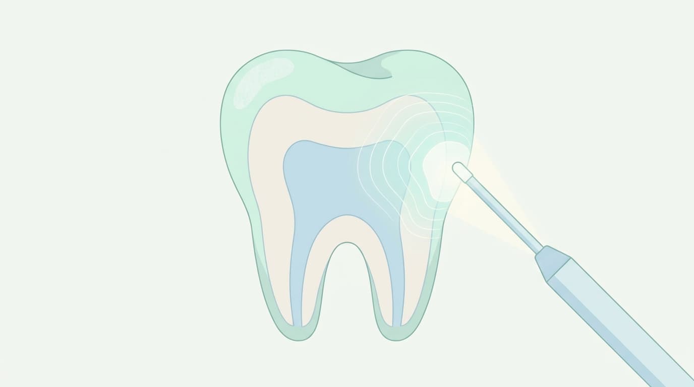
Resin infiltration targets enamel (the hard outer layer of the tooth) before a cavity forms on the surface, at the stage where minerals have already leached out and left behind a sponge-like network of microscopic pores. The classic example is white-spot lesions that appear on front teeth after orthodontic treatment. Unlike remineralization that refills a lesion with self-assembling peptides, resin infiltration works by lightly etching the tooth surface with acid to open a path into the microscopic pores, then flowing in a very low-viscosity resin to fill them. Once resin fills that space, it blocks acid and the acidic byproducts of bacteria from penetrating any further into the enamel.
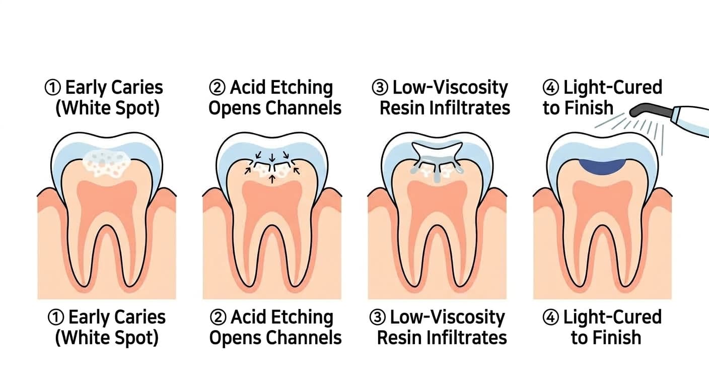
Several systematic reviews pooling clinical trials from multiple countries have repeatedly found that lesions treated with resin infiltration were about a third as likely to progress to cavities as untreated lesions.
The Same Duration Regardless of Lesion Depth
The problem is that this procedure has long followed a standardized protocol, etching and infiltrating for a fixed duration without regard to how deep each individual lesion actually is. Dentists have had no reliable way to check, by eye or on an X-ray, how deep a lesion runs inside the enamel, or whether etching and infiltration have actually reached that depth. Etching a shallow lesion for the standard duration removes more tooth structure than necessary, while stopping a deep lesion at the standard duration can leave the resin unable to fully penetrate, leaving the interior underfilled. This limited infiltration depth has long been cited as a limitation of resin infiltration.
Timing the Treatment by Seeing Inside with Light
A study published in September 2026 by a Chinese research team looked inside this process in real time using optical coherence tomography (OCT), an imaging technique that shines light into tissue and reconstructs internal structure from the reflected signal without cutting into it. By measuring how the brightness of light reflected from the enamel changed over time during acid etching and resin infiltration, the researchers plotted the relationship between etching and infiltration duration and the depth reached. Etching and infiltration depth increased over time but then plateaued, no longer deepening past a certain point.
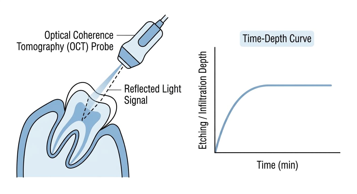
Based on this curve, the researchers designed a personalized protocol (OCT-IRI) that lengthens or shortens etching and infiltration time according to each lesion's actual depth, and pitted it against the conventional fixed-duration method (CRI), fluoride varnish (Duraphat), and no treatment at all.
Better in Shallow Lesions, Equal in Deep Lesions
The researchers created artificial lesions in extracted teeth, subjected them to renewed acid challenge, and compared how much secondary caries developed. In shallow lesions under 0.3 mm deep, the OCT-guided personalized approach produced markedly less secondary caries than either the fixed-duration conventional method or fluoride varnish. In deep lesions of 0.3 mm or more, however, there was no difference between the personalized approach and the conventional method, and both outperformed fluoride varnish. In deep lesions, fluoride varnish performed no better than no treatment at all.
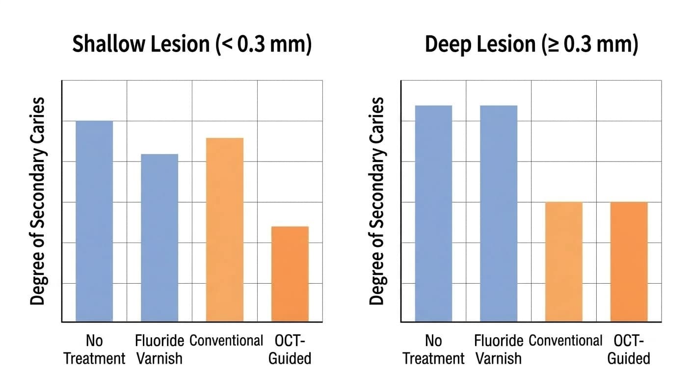
This is a preliminary study conducted only on extracted teeth with a small sample, so whether the same results hold inside an actual mouth still needs to be confirmed. Even so, it demonstrates a real possibility: a resin infiltration process that has always been standardized because there was no way to gauge lesion depth can instead be checked and adjusted in real time with light during the procedure. Resin infiltration cannot be used once a cavity has already formed; it applies only to early lesions that have not yet cavitated. Once a cavity forms, treatment must shift to restorations such as ion-releasing restorative materials or progression-arresting treatments such as SDF.