Question
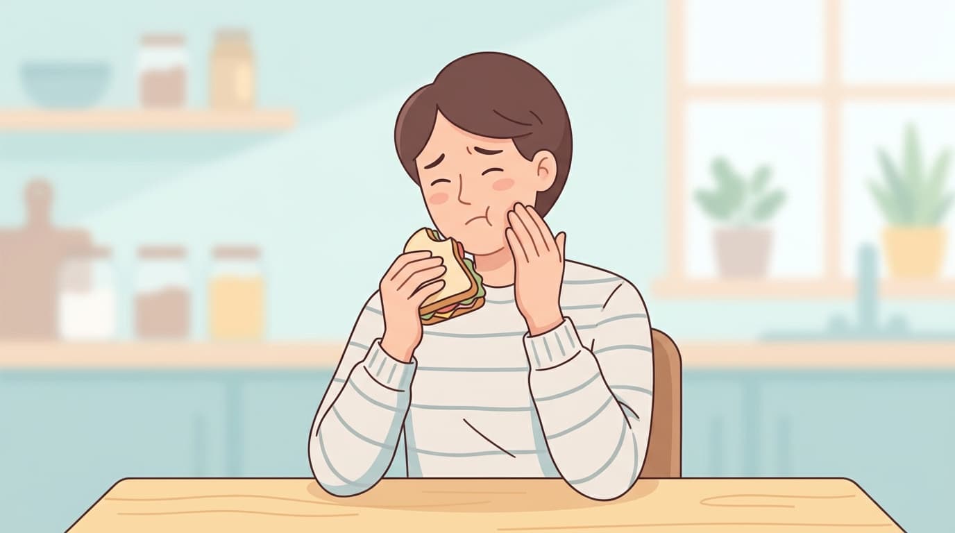
I feel a brief, sharp pain only when I bite down from one particular direction. Biting from the other side feels fine, but that sharp pain shoots through only when I bite in that one direction, and I'm worried it might mean my tooth has a crack.
Short Answer
A brief, sharp pain that appears only when biting from one particular direction is a commonly reported symptom of a cracked tooth (a tooth that hasn't broken completely but has a crack running along the chewing surface). However, this symptom alone isn't enough to confirm that a crack is present. A proper assessment requires testing each cusp (the pointed ridges of the chewing surface) individually to see whether biting force reproduces the pain, shining a light through the tooth to locate the crack, and checking whether the nerve is still alive. Even once a crack is confirmed, the treatment approach, ranging from observation to restoration, root canal treatment, or extraction, depends heavily on how far and how deep it has spread.
Why Does This Happen?
Why It Only Hurts When Biting From One Direction
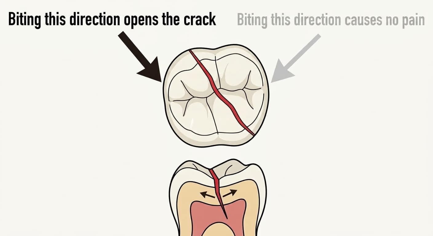
A cracked tooth has a fine, nearly vertical crack running inward from a narrow groove on the chewing surface. When biting force is applied or released, the two split segments shift slightly against each other, irritating the nerve tissue exposed at the crack, which produces a brief, sharp pain. This pain can occur both when biting down and when releasing the bite.
Because the crack runs in only one direction, the gap opens when force is applied along that direction but stays closed when biting from another direction, which is why pain appears selectively. This is especially common in teeth with large inlays or amalgam fillings covering most of the chewing surface, and in the molars of adults in their 30s and older.
What Tests Does the Dentist Perform?
When investigating the cause of pain while chewing, dentists don't rely on a single test to reach a conclusion; several tests are performed together.
The most basic test is the bite test. A fracture-detection device, cotton swab, or wooden wedge is placed on each cusp individually, and the dentist checks whether pain is reproduced when biting down and releasing. Identifying which cusp reproduces the pain helps narrow down where the crack is, and pain on biting or releasing raises the likelihood of both a crack and inflammation in the ligament surrounding the tooth.
Transillumination is also performed. Shining a strong fiber-optic light through the tooth causes the light to refract or reflect as it crosses the cracked surface, making that area appear darker. A dye-staining test may also be used to directly visualize the line the crack follows. Diagnostic accuracy improves the more of these tests are combined.
Testing also checks whether the nerve is still alive and what condition it's in. A brief tingling sensation to cold stimulation falls within the normal range, but an excessively sensitive reaction, or a dull pain that lingers for a while, signals that inflammation is progressing in the nerve, while no response at all suggests the nerve may have already died. Electric pulp testing is used for the same purpose. Gently tapping on the tooth also checks for inflammation in the surrounding ligament, and cracked teeth often react sensitively to this test as well. In the early stage, when the crack is limited to the dentin, an X-ray may show no noticeable change, so a normal-looking X-ray alone isn't taken to mean there's no problem.
Treatment Depends on the Crack's Location and Depth
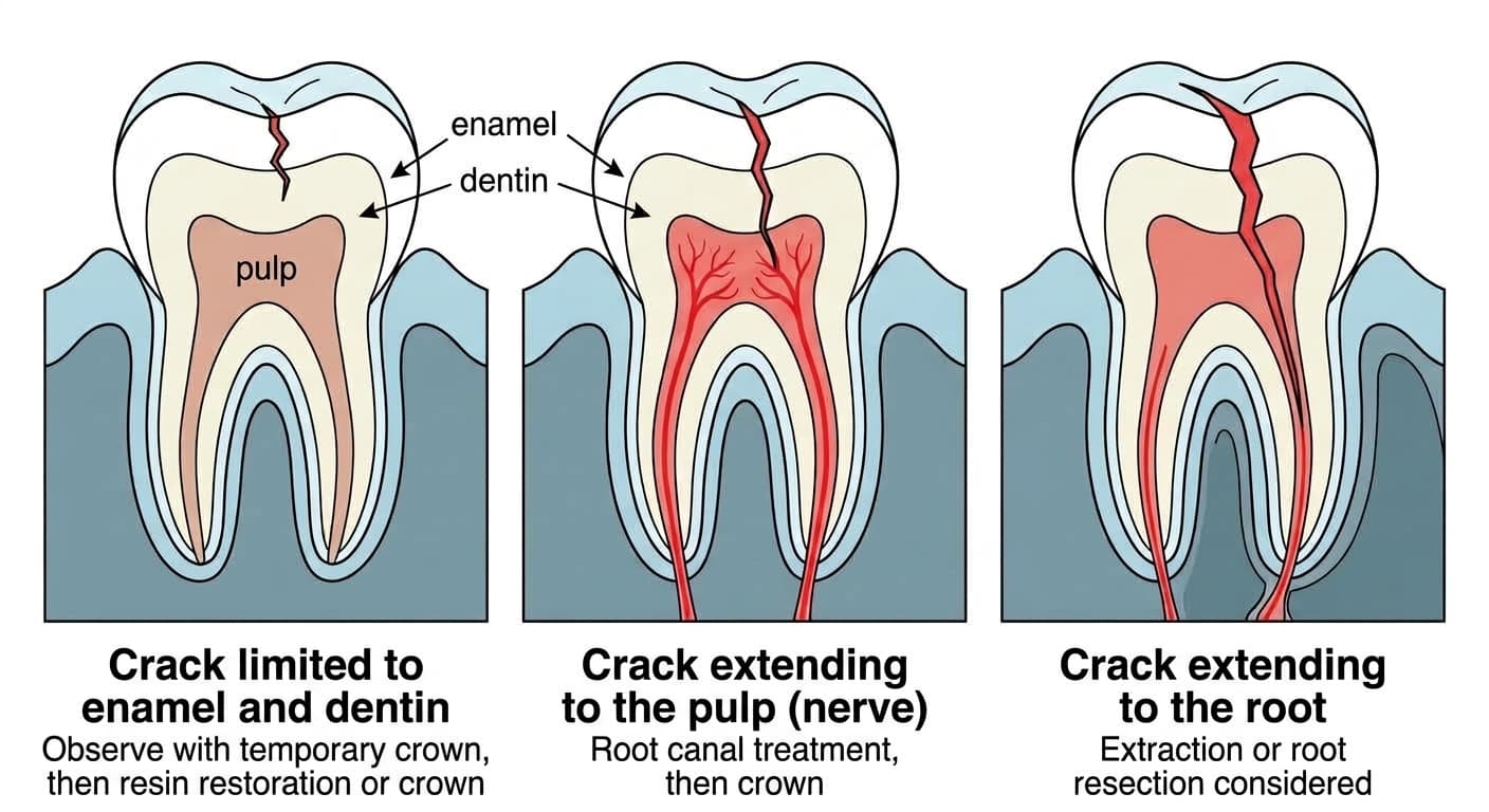
If the crack is still in its early stage, limited to the enamel or dentin surface, dentists often don't proceed straight to major treatment. Instead, the tooth is wrapped with an orthodontic band or temporary crown to see whether the symptoms settle. If the pain subsides, the tooth is finished either with a bonded composite resin restoration or a full crown to keep the crack from spreading further.
The situation changes if the crack extends to the pulp. Once irritation reaches inside the pulp chamber where the nerve sits, it can progress to pulpitis, which can bring on pain even at rest, and at this stage root canal treatment is needed. Because the crack remains in the tooth even after root canal treatment, the tooth is fully covered with a crown to keep biting force from being transmitted directly to the cracked area.
Rarely, the crack extends down to the root below the gumline. In this case, a narrow, deep gum pocket forms along the crack, an X-ray may show a distinctively shaped dark area around the root, and the inflammation can spread to the bone around the root tip, similar to apical periodontitis. A crack that has reached the root carries a poor prognosis, so extraction is generally the standard approach, but in a multi-rooted tooth such as a molar, if the bone supporting the remaining roots is in good condition, it may be possible to try resecting just the cracked root and saving the rest of the tooth. If the pain doesn't subside even after observation with a temporary crown, this situation should be suspected and the tooth should be reexamined.
If the Tooth Was Treated Recently, There May Be a Different Cause
If a tooth that recently had a filling or crown placed hurts only when biting from one particular direction, the cause may not be a crack but occlusal interference, where the restoration sits slightly higher than the surrounding teeth. In this case, simply adjusting the bite by smoothing down the high spot often resolves the pain within days to a few weeks.
That said, a reduction in pain doesn't necessarily mean things are improving. If the nerve dies from pulp necrosis, it stops responding to cold or electric stimulation entirely, which can feel like the pain has disappeared. If the brief pain while chewing keeps recurring, or if the pattern changes from an intermittent sharp twinge to throbbing pain even at rest, this shouldn't be left untreated, and a dental visit is needed.