Question
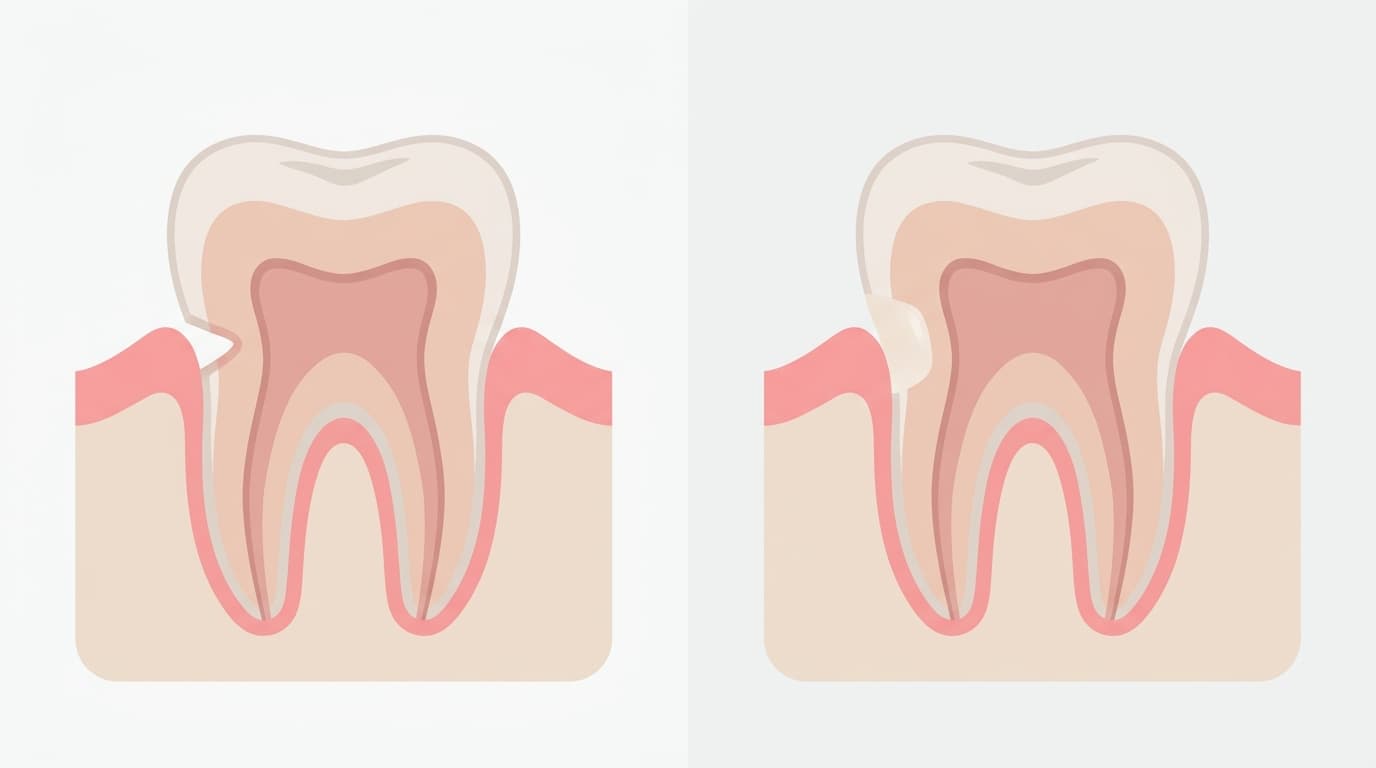
A common worry is that the area where the tooth meets the gum has become noticeably concave. It is not yet very sensitive, but looking in a mirror shows a visible notch, and the question is whether it should be filled with resin right away or can simply be watched.
Short Answer
The decision depends on the depth of the lesion, any symptoms, and the condition of the gum, considered together. If the lesion is shallow and there is no sensitivity or esthetic concern, it is often managed by watching it while addressing the habit that is causing it. If the lesion passes a certain depth, sensitivity does not settle with self-care, the gum is inflamed, or there is a risk of pulp exposure, restoring it with resin or a similar material is considered instead. When sensitivity is the only issue at this stage, the usual approach is to try calming it first with toothpaste or in-office treatment, and to move on to restoration only if that is not effective enough.
Why?
Why teeth notch near the gumline
In dentistry, this kind of lesion is called a non-carious cervical lesion, meaning that tooth structure at the neck of the tooth has been lost from a cause other than decay. It is not caused by a single factor but is a multifactorial lesion where several causes overlap. The main contributors are abrasion, where repeated friction such as toothbrushing wears down the tooth surface; erosion, where acids from foods or stomach acid chemically dissolve the tooth surface; and abfraction, where abnormal biting forces cause the tooth to flex slightly, concentrating stress at the neck of the tooth and chipping away structure in a wedge shape. As long as the underlying habit or irritant continues, the lesion tends to keep progressing.
When to watch and when to consider restoration
Textbooks describe evaluating several factors together when deciding whether to restore a lesion. These include the depth of the lesion, whether active decay is also present, whether the appearance is an esthetic concern, whether there is sensitivity (dentin hypersensitivity), whether the gum is inflamed or further recession needs to be prevented, and whether there is a risk of exposing the pulp (the tooth's nerve tissue). If none of these apply, the lesion is often simply watched while managing the underlying habit, rather than restored right away.
Depth is also given as a specific benchmark. Textbooks list a lesion depth greater than 1.5mm, or by another standard, an axial wall depth greater than 2mm, as a threshold for considering restoration. However, depth alone does not decide the matter; whether the other factors above are also present is considered together.
Sensitivity is addressed step by step
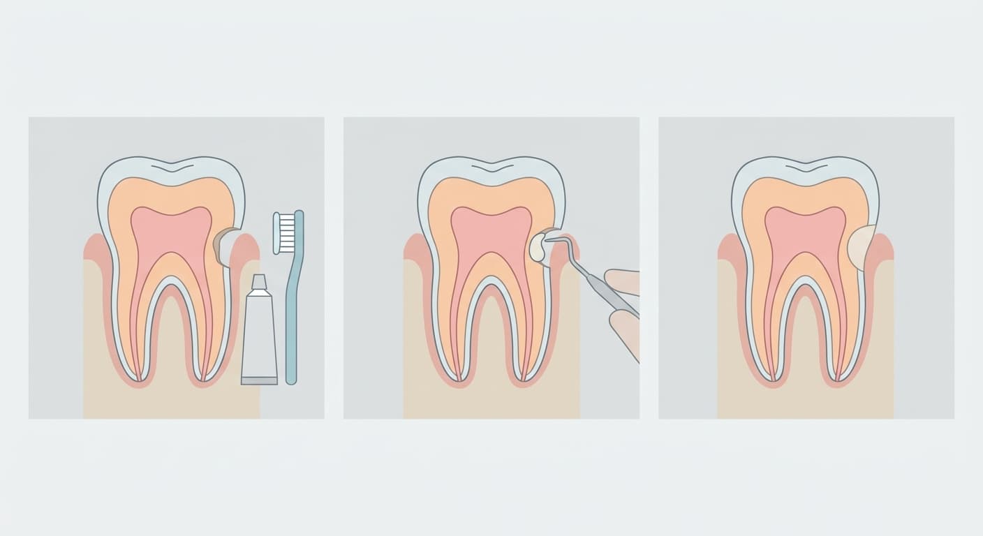
When a lesion is accompanied by sensitivity, the symptom itself is often addressed first. At home, this usually means using a desensitizing toothpaste that calms nerve excitability or contains minerals that refill the openings of the dentinal tubules, used consistently over time. If that is not enough, in-office treatments such as tubule-sealing agents, fluoride varnish, or laser treatment can be done at the dental clinic. Only if symptoms still do not settle enough after these steps, and other factors such as lesion depth, esthetics, or the risk of pulp exposure are also present, is restoring the exposed surface with a material such as resin or glass ionomer considered. This stepwise approach is covered in more detail in the article on dentin hypersensitivity.
When gum condition is also involved
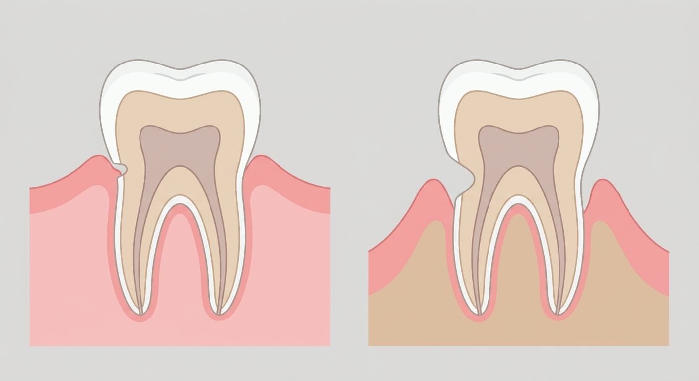
Some cases involve gum recession alongside the lesion. If the tissue covering the gum (attached gingiva) is insufficient, or if Miller Class I or Class II gingival recession is also present, a combined approach involving a graft to raise the gum tissue along with a resin-modified glass ionomer restoration may be considered. In these cases, grafting only the area where enamel is exposed is not recommended, since gum tissue is unlikely to attach and gain blood supply there. A combined approach that restores the defect at the same time is usually discussed instead.
When abfraction from pronounced flexural forces on the tooth is the cause, one more factor comes into play when choosing a material. A microfilled composite resin or resin-modified glass ionomer with a low elastic modulus, which can flex slightly along with the tooth, is recommended. More detailed criteria for choosing between composite resin and glass ionomer depending on the location of the lesion and the condition of the gum are covered in Should a Notched Tooth Neck Be Filled With Resin or Glass Ionomer?.